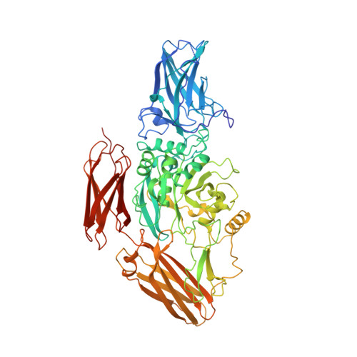
1GGT

THREE-DIMENSIONAL STRUCTURE OF A TRANSGLUTAMINASE: HUMAN BLOOD COAGULATION FACTOR XIII


Experimental Data Snapshot

  • Method: X-RAY DIFFRACTION
  • Resolution: 2.65 Å
  • R-Value Work: 0.216 
  • R-Value Observed: 0.216 

wwPDB Validation   3D Report Full Report


This is version 1.4 of the entry. See complete history


Literature

Three-dimensional structure of a transglutaminase: human blood coagulation factor XIII.

Yee, V.C.Pedersen, L.C.Le Trong, I.Bishop, P.D.Stenkamp, R.E.Teller, D.C.

(1994) Proc Natl Acad Sci U S A 91: 7296-7300

  • DOI: https://doi.org/10.1073/pnas.91.15.7296
  • Primary Citation of Related Structures:  
    1GGT

  • PubMed Abstract: 

    Mechanical stability in many biological materials is provided by the crosslinking of large structural proteins with gamma-glutamyl-epsilon-lysyl amide bonds. The three-dimensional structure of human recombinant factor XIII (EC 2.3.2.13 zymogen; protein-glutamine:amine gamma-glutamyltransferase a chain), a transglutaminase zymogen, has been solved at 2.8-A resolution by x-ray crystallography. This structure shows that each chain of the homodimeric protein is folded into four sequential domains. A catalytic triad reminiscent of that observed in cysteine proteases has been identified in the core domain. The amino-terminal activation peptide of each subunit crosses the dimer interface and partially occludes the opening of the catalytic cavity in the second subunit, preventing substrate binding to the zymogen. A proposal for the mechanism of activation by thrombin and calcium is made that details the structural events leading to active factor XIIIa'.


  • Organizational Affiliation

    Department of Biochemistry, University of Washington, Seattle 98195.


Macromolecules
Find similar proteins by:  (by identity cutoff)  |  3D Structure
Entity ID: 1
MoleculeChains Sequence LengthOrganismDetailsImage
COAGULATION FACTOR XIII
A, B
731Homo sapiensMutation(s): 0 
EC: 2.3.2.13
UniProt & NIH Common Fund Data Resources
Find proteins for P00488 (Homo sapiens)
Explore P00488 
Go to UniProtKB:  P00488
PHAROS:  P00488
GTEx:  ENSG00000124491 
Entity Groups  
Sequence Clusters30% Identity50% Identity70% Identity90% Identity95% Identity100% Identity
UniProt GroupP00488
Sequence Annotations
Expand
  • Reference Sequence
Experimental Data & Validation

Experimental Data

  • Method: X-RAY DIFFRACTION
  • Resolution: 2.65 Å
  • R-Value Work: 0.216 
  • R-Value Observed: 0.216 
  • Space Group: P 21 21 2
Unit Cell:
Length ( Å )Angle ( ˚ )
a = 101.2α = 90
b = 182.7β = 90
c = 93.4γ = 90
Software Package:
Software NamePurpose
X-PLORmodel building
X-PLORrefinement
X-PLORphasing

Structure Validation

View Full Validation Report


Entry History 

Deposition Data

Revision History  (Full details and data files)

  • Version 1.0: 1995-07-31
    Type: Initial release
  • Version 1.1: 2008-03-03
    Changes: Version format compliance
  • Version 1.2: 2011-07-13
    Changes: Version format compliance
  • Version 1.3: 2017-11-29
    Changes: Derived calculations, Other
  • Version 1.4: 2024-02-07
    Changes: Data collection, Database references