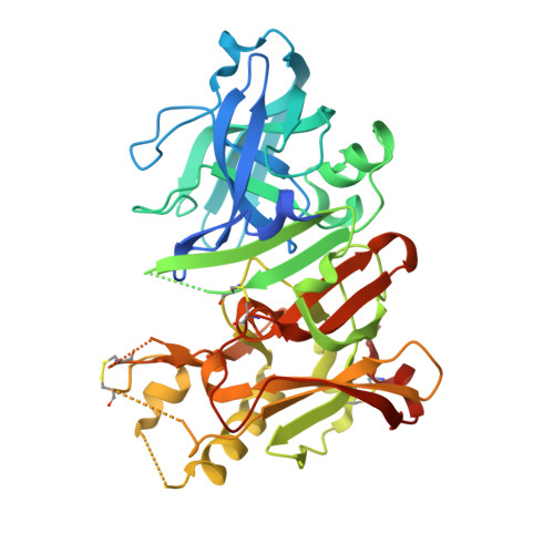
3VV8

Crystal structure of beta secetase in complex with 2-amino-3-methyl-6-((1S,2R)-2-(3'-methylbiphenyl-4-yl)cyclopropyl)pyrimidin-4(3H)-one


Experimental Data Snapshot

  • Method: X-RAY DIFFRACTION
  • Resolution: 2.50 Å
  • R-Value Free: 0.287 
  • R-Value Work: 0.224 
  • R-Value Observed: 0.227 

wwPDB Validation   3D Report Full Report


Ligand Structure Quality Assessment 


This is version 1.2 of the entry. See complete history


Literature

Conformational restriction approach to beta-secretase (BACE1) inhibitors: effect of a cyclopropane ring to induce an alternative binding mode.

Yonezawa, S.Yamamoto, T.Yamakawa, H.Muto, C.Hosono, M.Hattori, K.Higashino, K.Yutsudo, T.Iwamoto, H.Kondo, Y.Sakagami, M.Togame, H.Tanaka, Y.Nakano, T.Takemoto, H.Arisawa, M.Shuto, S.

(2012) J Med Chem 55: 8838-8858

  • DOI: https://doi.org/10.1021/jm3011405
  • Primary Citation of Related Structures:  
    3VV6, 3VV7, 3VV8

  • PubMed Abstract: 

    Improvement of a drug's binding activity using the conformational restriction approach with sp³ hybridized carbon is becoming a key strategy in drug discovery. We applied this approach to BACE1 inhibitors and designed four stereoisomeric cyclopropane compounds in which the ethylene linker of a known amidine-type inhibitor 2 was replaced with chiral cyclopropane rings. The synthesis and biologic evaluation of these compounds revealed that the cis-(1S,2R) isomer 6 exhibited the most potent BACE1 inhibitory activity among them. X-ray structure analysis of the complex of 6 and BACE1 revealed that its unique binding mode is due to the apparent CH-π interaction between the rigid cyclopropane ring and the Tyr71 side chain. A derivatization study using 6 as a lead molecule led to the development of highly potent inhibitors in which the structure-activity relationship as well as the binding mode of the compounds clearly differ from those of known amidine-type inhibitors.


  • Organizational Affiliation

    Shionogi Innovation Center for Drug Discovery, Shionogi & Co., Ltd., Kita-21 Nishi-11 Kita-ku, Sapporo 001-0021, Japan. [email protected]


Macromolecules
Find similar proteins by:  (by identity cutoff)  |  3D Structure
Entity ID: 1
MoleculeChains Sequence LengthOrganismDetailsImage
Beta-secretase 1416Homo sapiensMutation(s): 0 
Gene Names: BACE1
EC: 3.4.23.46
UniProt & NIH Common Fund Data Resources
Find proteins for P56817 (Homo sapiens)
Explore P56817 
Go to UniProtKB:  P56817
PHAROS:  P56817
GTEx:  ENSG00000186318 
Entity Groups  
Sequence Clusters30% Identity50% Identity70% Identity90% Identity95% Identity100% Identity
UniProt GroupP56817
Sequence Annotations
Expand
  • Reference Sequence
Small Molecules
Ligands 2 Unique
IDChains Name / Formula / InChI Key2D Diagram3D Interactions
B02
Query on B02

Download Ideal Coordinates CCD File 
C [auth A]2-amino-3-methyl-6-[(1S,2R)-2-(3'-methylbiphenyl-4-yl)cyclopropyl]pyrimidin-4(3H)-one
C21 H21 N3 O
STUBOTPNCNDBOA-ROUUACIJSA-N
GOL
Query on GOL

Download Ideal Coordinates CCD File 
B [auth A]GLYCEROL
C3 H8 O3
PEDCQBHIVMGVHV-UHFFFAOYSA-N
Binding Affinity Annotations 
IDSourceBinding Affinity
B02 PDBBind:  3VV8 IC50: 1.71e+4 (nM) from 1 assay(s)
BindingDB:  3VV8 IC50: 1.71e+4 (nM) from 1 assay(s)
Experimental Data & Validation

Experimental Data

  • Method: X-RAY DIFFRACTION
  • Resolution: 2.50 Å
  • R-Value Free: 0.287 
  • R-Value Work: 0.224 
  • R-Value Observed: 0.227 
  • Space Group: P 61 2 2
Unit Cell:
Length ( Å )Angle ( ˚ )
a = 101.92α = 90
b = 101.92β = 90
c = 169.26γ = 120
Software Package:
Software NamePurpose
MOSFLMdata reduction
SCALAdata scaling
MOLREPphasing
REFMACrefinement
PDB_EXTRACTdata extraction

Structure Validation

View Full Validation Report


Ligand Structure Quality Assessment 


Entry History 

Revision History  (Full details and data files)

  • Version 1.0: 2012-10-24
    Type: Initial release
  • Version 1.1: 2013-09-04
    Changes: Database references
  • Version 1.2: 2024-11-20
    Changes: Data collection, Database references, Derived calculations, Structure summary