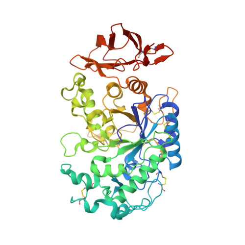
Directed "in situ" inhibitor elongation as a strategy to structurally characterize the covalent glycosyl-enzyme intermediate of human pancreatic alpha-amylase
Zhang, R., Li, C., Williams, L.K., Rempel, B.P., Brayer, G.D., Withers, S.G.(2009) Biochemistry 48: 10752-10764
- PubMed: 19803533
- DOI: https://doi.org/10.1021/bi901400p
- Primary Citation of Related Structures:
3IJ7, 3IJ8, 3IJ9 - PubMed Abstract:
While covalent catalytic intermediates of retaining alpha-transglycosylases have been structurally characterized previously, no such information for a hydrolytic alpha-amylase has been obtained. This study presents a new "in situ" enzymatic elongation methodology that, for the first time, has allowed the isolation and structural characterization of a catalytically competent covalent glycosyl-enzyme intermediate with human pancreatic alpha-amylase. This has been achieved by the use of a 5-fluoro-beta-l-idosyl fluoride "warhead" in conjunction with either alpha-maltotriosyl fluoride or 4'-O-methyl-alpha-maltosyl fluoride as elongation agents. This generates an oligosaccharyl-5-fluoroglycosyl fluoride that then reacts with the free enzyme. The resultant covalent intermediates are extremely stable, with hydrolytic half-lives on the order of 240 h for the trisaccharide complex. In the presence of maltose, however, they undergo turnover via transglycosylation according to a half-life of less than 1 h. Structural studies of intermediate complexes unambiguously show the covalent attachment of a 5-fluoro-alpha-l-idosyl moiety in the chair conformation to the side chain of the catalytic nucleophile D197. The elongated portions of the intermediate complexes are found to bind in the high-affinity -2 and -3 binding subsites, forming extensive hydrogen-bonding interactions. Comparative structural analyses with the related noncovalent complex formed by acarbose highlight the structural rigidity of the enzyme surface during catalysis and the key role that substrate conformational flexibility must play in this process. Taken together, the structural data provide atomic details of several key catalytic steps. The scope of this elongation approach to probe the active sites and catalytic mechanisms of alpha-amylases is further demonstrated through preliminary experiments with porcine pancreatic alpha-amylase.
Organizational Affiliation:
Department of Chemistry, University of British Columbia, Vancouver, British Columbia V6T 1Z1, Canada.


