4Q30

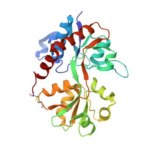
Nitrowillardiine bound to the ligand binding domain of GluA2 at pH 3.5


Experimental Data Snapshot

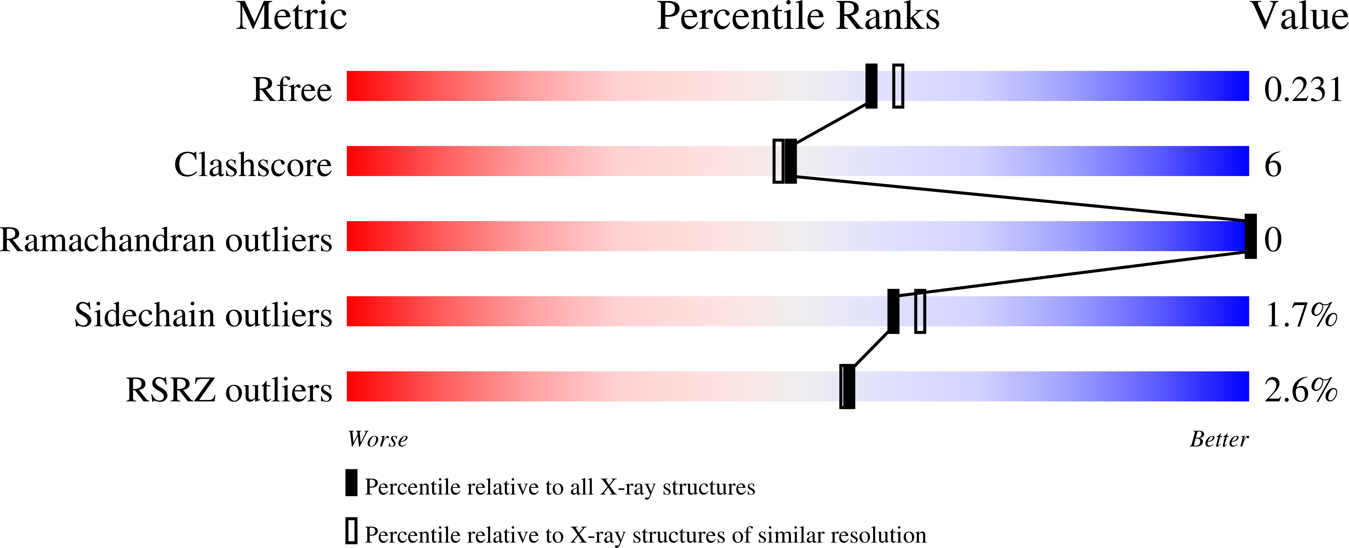
  • Method: X-RAY DIFFRACTION
  • Resolution: 2.03 Å
  • R-Value Free: 0.230 
  • R-Value Work: 0.186 
  • R-Value Observed: 0.187 

Starting Model: experimental
View more details

wwPDB Validation   3D Report Full Report


Ligand Structure Quality Assessment 


This is version 1.4 of the entry. See complete history


Literature

Thermodynamics and mechanism of the interaction of willardiine partial agonists with a glutamate receptor: implications for drug development.

Martinez, M.Ahmed, A.H.Loh, A.P.Oswald, R.E.

(2014) Biochemistry 53: 3790-3795

  • DOI: https://doi.org/10.1021/bi500511m
  • Primary Citation of Related Structures:  
    4Q30

  • PubMed Abstract: 

    Understanding the thermodynamics of binding of a lead compound to a receptor can provide valuable information for drug design. The binding of compounds, particularly partial agonists, to subtypes of the α-amino-3-hydroxy-5-methyl-4-isoxazole-propionic acid (AMPA) receptor is, in some cases, driven by increases in entropy. Using a series of partial agonists based on the structure of the natural product, willardiine, we show that the charged state of the ligand determines the enthalpic contribution to binding. Willardiines have uracil rings with pKa values ranging from 5.5 to 10. The binding of the charged form is largely driven by enthalpy, while that of the uncharged form is largely driven by entropy. This is due at least in part to changes in the hydrogen bonding network within the binding site involving one water molecule. This work illustrates the importance of charge to the thermodynamics of binding of agonists and antagonists to AMPA receptors and provides clues for further drug discovery.


  • Organizational Affiliation

    Department of Molecular Medicine, Cornell University , Ithaca, New York 14853, United States.


Macromolecules
Find similar proteins by:  (by identity cutoff)  |  3D Structure
Entity ID: 1
MoleculeChains Sequence LengthOrganismDetailsImage
Glutamate receptor 2 CHIMERIC PROTEINA [auth B],
B [auth D],
C [auth F]
258Rattus norvegicusMutation(s): 0 
Gene Names: Glur2Gria2
UniProt
Find proteins for P19491 (Rattus norvegicus)
Explore P19491 
Go to UniProtKB:  P19491
Entity Groups  
Sequence Clusters30% Identity50% Identity70% Identity90% Identity95% Identity100% Identity
UniProt GroupP19491
Sequence Annotations
Expand
  • Reference Sequence
Small Molecules
Ligands 2 Unique
IDChains Name / Formula / InChI Key2D Diagram3D Interactions
NWD
Query on NWD

Download Ideal Coordinates CCD File 
D [auth B],
G [auth D],
J [auth F]
3-(5-nitro-2,4-dioxo-3,4-dihydropyrimidin-1(2H)-yl)-L-alanine
C7 H8 N4 O6
IEBVITXSHAFLJR-VKHMYHEASA-N
ZN
Query on ZN

Download Ideal Coordinates CCD File 
E [auth B],
F [auth B],
H [auth D],
I [auth D],
K [auth F]
ZINC ION
Zn
PTFCDOFLOPIGGS-UHFFFAOYSA-N
Experimental Data & Validation

Experimental Data

  • Method: X-RAY DIFFRACTION
  • Resolution: 2.03 Å
  • R-Value Free: 0.230 
  • R-Value Work: 0.186 
  • R-Value Observed: 0.187 
  • Space Group: P 2 21 21
Unit Cell:
Length ( Å )Angle ( ˚ )
a = 47.797α = 90
b = 113.27β = 90
c = 164.321γ = 90
Software Package:
Software NamePurpose
HKL-2000data collection
PHENIXmodel building
PHENIXrefinement
HKL-2000data reduction
HKL-2000data scaling
PHENIXphasing

Structure Validation

View Full Validation Report


Ligand Structure Quality Assessment 


Entry History 

Deposition Data

Revision History  (Full details and data files)

  • Version 1.0: 2014-06-04
    Type: Initial release
  • Version 1.1: 2014-07-02
    Changes: Database references
  • Version 1.2: 2017-07-26
    Changes: Data collection, Source and taxonomy
  • Version 1.3: 2023-09-20
    Changes: Data collection, Database references, Derived calculations, Refinement description
  • Version 1.4: 2024-11-20
    Changes: Structure summary