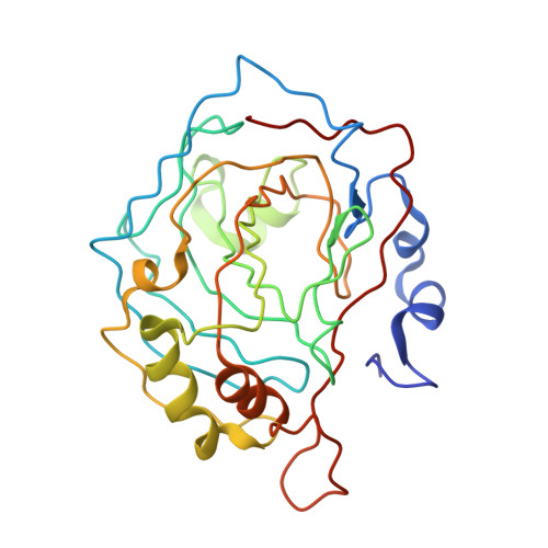
1CIN

THE POSITIONS OF HIS-64 AND A BOUND WATER IN HUMAN CARBONIC ANHYDRASE II UPON BINDING THREE STRUCTURALLY RELATED INHIBITORS


Experimental Data Snapshot

  • Method: X-RAY DIFFRACTION
  • Resolution: 2.10 Å

wwPDB Validation   3D Report Full Report


This is version 1.3 of the entry. See complete history


Literature

Positions of His-64 and a bound water in human carbonic anhydrase II upon binding three structurally related inhibitors.

Smith, G.M.Alexander, R.S.Christianson, D.W.McKeever, B.M.Ponticello, G.S.Springer, J.P.Randall, W.C.Baldwin, J.J.Habecker, C.N.

(1994) Protein Sci 3: 118-125

  • DOI: https://doi.org/10.1002/pro.5560030115
  • Primary Citation of Related Structures:  
    1CIL, 1CIM, 1CIN

  • PubMed Abstract: 

    The 3-dimensional structure of human carbonic anhydrase II (HCAII; EC 4.2.1.1) complexed with 3 structurally related inhibitors, 1a, 1b, and 1c, has been determined by X-ray crystallographic methods. The 3 inhibitors (1a = C8H12N2O4S3) vary only in the length of the substituent on the 4-amino group: 1a, proton; 1b, methyl; and 1c, ethyl. The binding constants (Ki's) for 1a, 1b, and 1c to HCAII are 1.52, 1.88, and 0.37 nM, respectively. These structures were solved to learn if any structural cause could be found for the difference in binding. In the complex with inhibitors 1a and 1b, electron density can be observed for His-64 and a bound water molecule in the native positions. When inhibitor 1c is bound, the side chain attached to the 4-amino group is positioned so that His-64 can only occupy the alternate position and the bound water is absent. While a variety of factors contribute to the observed binding constants, the major reason 1c binds tighter to HCAII than does 1a or 1b appears to be entropy: the increase in entropy when the bound water molecule is released contributes to the increase in binding and overcomes the small penalty for putting the His-64 side chain in a higher energy state.


  • Organizational Affiliation

    Merck Research Laboratories, West Point, Pennsylvania 19486.


Macromolecules
Find similar proteins by:  (by identity cutoff)  |  3D Structure
Entity ID: 1
MoleculeChains Sequence LengthOrganismDetailsImage
CARBONIC ANHYDRASE II259Homo sapiensMutation(s): 0 
EC: 4.2.1.1 (PDB Primary Data), 4.2.1.69 (UniProt)
UniProt & NIH Common Fund Data Resources
Find proteins for P00918 (Homo sapiens)
Explore P00918 
Go to UniProtKB:  P00918
PHAROS:  P00918
GTEx:  ENSG00000104267 
Entity Groups  
Sequence Clusters30% Identity50% Identity70% Identity90% Identity95% Identity100% Identity
UniProt GroupP00918
Sequence Annotations
Expand
  • Reference Sequence
Small Molecules
Ligands 3 Unique
IDChains Name / Formula / InChI Key2D Diagram3D Interactions
MTS
Query on MTS

Download Ideal Coordinates CCD File 
D [auth A](4S-TRANS)-4-(METHYLAMINO)-5,6-DIHYDRO-6-METHYL-4H-THIENO(2,3-B)THIOPYRAN-2-SULFONAMIDE-7,7-DIOXIDE
C9 H14 N2 O4 S3
PYXFWOIZPYXNRU-FSPLSTOPSA-N
MMC
Query on MMC

Download Ideal Coordinates CCD File 
C [auth A]METHYL MERCURY ION
C H3 Hg
DBUXSCUEGJMZAE-UHFFFAOYSA-N
ZN
Query on ZN

Download Ideal Coordinates CCD File 
B [auth A]ZINC ION
Zn
PTFCDOFLOPIGGS-UHFFFAOYSA-N
Binding Affinity Annotations 
IDSourceBinding Affinity
MTS PDBBind:  1CIN Ki: 1.88 (nM) from 1 assay(s)
Experimental Data & Validation

Experimental Data

  • Method: X-RAY DIFFRACTION
  • Resolution: 2.10 Å
  • Space Group: P 1 21 1
Unit Cell:
Length ( Å )Angle ( ˚ )
a = 42.83α = 90
b = 42.04β = 104.78
c = 72.62γ = 90
Software Package:
Software NamePurpose
X-PLORmodel building
X-PLORrefinement
X-PLORphasing

Structure Validation

View Full Validation Report


Entry History 

Revision History  (Full details and data files)

  • Version 1.0: 1994-01-31
    Type: Initial release
  • Version 1.1: 2008-03-24
    Changes: Version format compliance
  • Version 1.2: 2011-07-13
    Changes: Version format compliance
  • Version 1.3: 2024-02-07
    Changes: Data collection, Database references, Derived calculations, Other