1GA0

STRUCTURE OF THE E. CLOACAE GC1 BETA-LACTAMASE WITH A CEPHALOSPORIN SULFONE INHIBITOR


Experimental Data Snapshot

  • Method: X-RAY DIFFRACTION
  • Resolution: 1.60 Å
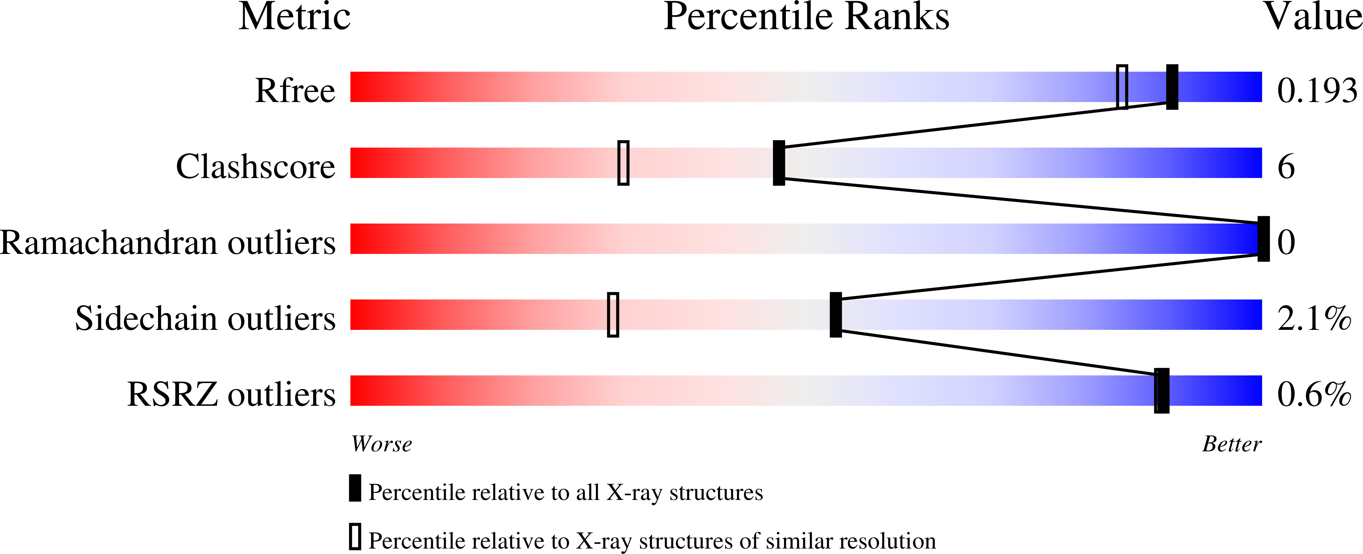
  • R-Value Free: 0.208 
  • R-Value Work: 0.181 
  • R-Value Observed: 0.183 

Starting Model: experimental
View more details

wwPDB Validation   3D Report Full Report


Ligand Structure Quality Assessment 


This is version 1.5 of the entry. See complete history


Literature

Inhibition of class C beta-lactamases: structure of a reaction intermediate with a cephem sulfone.

Crichlow, G.V.Nukaga, M.Doppalapudi, V.R.Buynak, J.D.Knox, J.R.

(2001) Biochemistry 40: 6233-6239

  • DOI: https://doi.org/10.1021/bi010131s
  • Primary Citation of Related Structures:  
    1GA0

  • PubMed Abstract: 

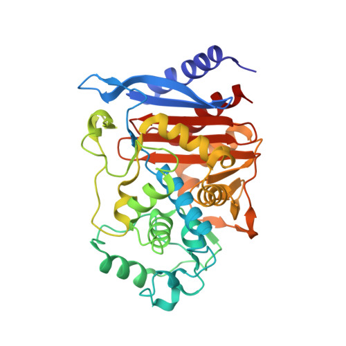
    The crystallographic structure of the Enterobacter cloacae GC1 extended-spectrum class C beta-lactamase, inhibited by a new 7-alkylidenecephalosporin sulfone, has been determined by X-ray diffraction at 100 K to a resolution of 1.6 A. The crystal structure was solved by molecular replacement using the unliganded structure [Crichlow et al. (1999) Biochemistry 38, 10256-10261] and refined to a crystallographic R-factor equal to 0.183 (R(free) 0.208). Cryoquenching of the reaction of the sulfone with the enzyme produced an intermediate that is covalently bound via Ser64. After acylation of the beta-lactam ring, the dihydrothiazine dioxide ring opened with departure of the sulfinate. Nucleophilic attack of a side chain pyridine nitrogen atom on the C6 atom of the resultant imine yielded a bicyclic aromatic system which helps to stabilize the acyl enzyme to hydrolysis. A structural assist to this resonance stabilization is the positioning of the anionic sulfinate group between the probable catalytic base (Tyr150) and the acyl ester bond so as to block the approach of a potentially deacylating water molecule. Comparison of the liganded and unliganded protein structures showed that a major movement (up to 7 A) and refolding of part of the Omega-loop (215-224) accompanies the binding of the inhibitor. This conformational flexibility in the Omega-loop may form the basis of an extended-spectrum activity of class C beta-lactamases against modern cephalosporins.


  • Organizational Affiliation

    Department of Molecular and Cell Biology, The University of Connecticut, Storrs, Connecticut 06269-3125, USA.


Macromolecules
Find similar proteins by:  (by identity cutoff)  |  3D Structure
Entity ID: 1
MoleculeChains Sequence LengthOrganismDetailsImage
BETA-LACTAMASE364Enterobacter cloacaeMutation(s): 0 
Gene Names: BLAC
EC: 3.5.2.6
UniProt
Find proteins for Q59401 (Enterobacter cloacae)
Explore Q59401 
Go to UniProtKB:  Q59401
Entity Groups  
Sequence Clusters30% Identity50% Identity70% Identity90% Identity95% Identity100% Identity
UniProt GroupQ59401
Sequence Annotations
Expand
  • Reference Sequence
Experimental Data & Validation

Experimental Data

  • Method: X-RAY DIFFRACTION
  • Resolution: 1.60 Å
  • R-Value Free: 0.208 
  • R-Value Work: 0.181 
  • R-Value Observed: 0.183 
  • Space Group: P 21 21 2
Unit Cell:
Length ( Å )Angle ( ˚ )
a = 77α = 90
b = 69β = 90
c = 62.5γ = 90
Software Package:
Software NamePurpose
AMoREphasing
CNSrefinement
DENZOdata reduction
SCALEPACKdata scaling

Structure Validation

View Full Validation Report


Ligand Structure Quality Assessment 


Entry History 

Deposition Data

Revision History  (Full details and data files)

  • Version 1.0: 2001-06-06
    Type: Initial release
  • Version 1.1: 2008-04-27
    Changes: Version format compliance
  • Version 1.2: 2011-07-13
    Changes: Non-polymer description, Version format compliance
  • Version 1.3: 2011-11-16
    Changes: Atomic model
  • Version 1.4: 2023-08-09
    Changes: Data collection, Database references, Derived calculations, Refinement description
  • Version 1.5: 2024-11-13
    Changes: Structure summary