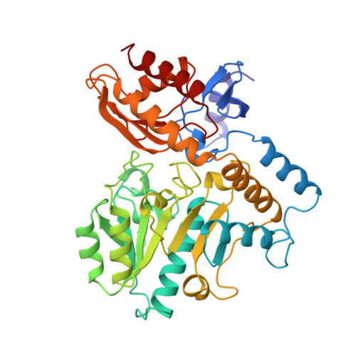
Human ornithine aminotransferase complexed with L-canaline and gabaculine: structural basis for substrate recognition.
Shah, S.A., Shen, B.W., Brunger, A.T.(1997) Structure 5: 1067-1075
- PubMed: 9309222
- DOI: https://doi.org/10.1016/s0969-2126(97)00258-x
- Primary Citation of Related Structures:
1GBN, 2CAN - PubMed Abstract:
Ornithine aminotransferase (OAT) is a 45 kDa pyridoxal-5'-phosphate (PLP)-dependent enzyme that catalyzes the conversion of L-ornithine and 2-oxoglutarate to glutamate-delta-semialdehyde and glutamic acid, respectively. In humans, loss of OAT function causes an accumulation of ornithine that results in gyrate atrophy of the choroid and retina, a disease that progressively leads to blindness. In an effort to learn more about the structural basis of this enzyme's function, we have determined the X-ray structures of OAT in complex with two enzyme-activated suicide substrates: L-canaline, an ornithine analog, and gabaculine, an irreversible inhibitor of several related aminotransferases. The structures of human OAT bound to the inhibitors gabaculine and L-canaline were solved to 2.3 A at 110K by difference Fourier techniques. Both inhibitors coordinate similarly in the active site, binding covalently to the PLP cofactor and causing a 20 degrees rotation in the cofactor tilt relative to the ligand-free form. Aromatic-aromatic interactions occur between the bound gabaculine molecule and active-site residues Tyr85 and Phe177, whereas Tyr55 and Arg180 provide specific contacts to the alpha-amino and carboxyl groups of L-canaline. The OAT-L-canaline complex structure implicates Tyr55 and Arg180 as the residues involved in coordinating with the natural substrate ornithine during normal enzyme turnover. This correlates well with two enzyme-inactivating point mutations associated with gyrate atrophy, Tyr55-->His and Arg180-->Thr. The OAT-gabaculine complex provides the first structural evidence that the potency of the inhibitor is due to energetically favourable aromatic interactions with residues in the active site. This aromatic-binding mode may be relevant to structure-based drug design efforts against other omega-aminotransferase targets, such as GABA aminotransferase.
Organizational Affiliation:
Department of Molecular Biophysics and Biochemistry, Yale University, New Haven, CT 06520, USA.


