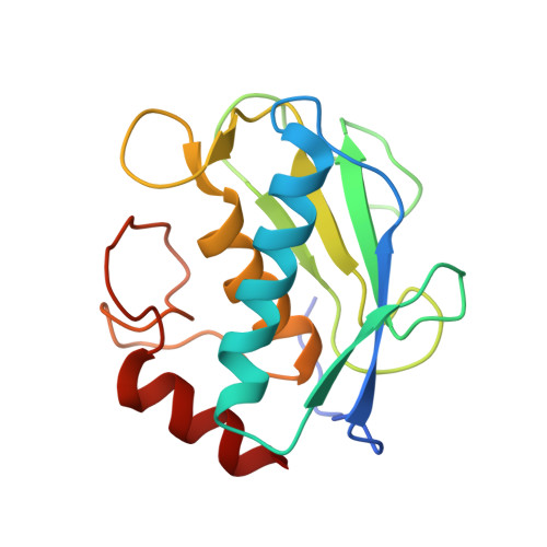
1GKC

MMP9-inhibitor complex


Experimental Data Snapshot

  • Method: X-RAY DIFFRACTION
  • Resolution: 2.30 Å
  • R-Value Free: 0.239 
  • R-Value Work: 0.207 

Starting Model: experimental
View more details

wwPDB Validation   3D Report Full Report


Ligand Structure Quality Assessment 


This is version 1.2 of the entry. See complete history


Literature

Crystal Structure of Mmp9 in Complex with a Reverse Hydroxamate Inhibitor

Rowsell, S.Hawtin, P.Minshull, C.A.Jepson, H.Brockbank, S.Barratt, D.Slater, A.M.Mcpheat, W.Waterson, D.Henney, A.Pauptit, R.A.

(2002) J Mol Biol 319: 173

  • DOI: https://doi.org/10.1016/S0022-2836(02)00262-0
  • Primary Citation of Related Structures:  
    1GKC, 1GKD

  • PubMed Abstract: 

    Matrix metalloproteinases (MMPs) and their inhibitors are important in connective tissue re-modelling in diseases of the cardiovascular system, such as atherosclerosis. Various members of the MMP family have been shown to be expressed in atherosclerotic lesions, but MMP9 is consistently seen in inflammatory atherosclerotic lesions. MMP9 over-expression is implicated in the vascular re-modelling events preceding plaque rupture (the most common cause of acute myocardial infarction). Reduced MMP9 activity, either by genetic manipulation or through pharmacological intervention, has an impact on ventricular re-modelling following infarction. MMP9 activity may therefore represent a key mechanism in the pathogenesis of heart failure. We have determined the crystal structure, at 2.3 A resolution, of the catalytic domain of human MMP9 bound to a peptidic reverse hydroxamate inhibitor as well as the complex of the same inhibitor bound to an active-site mutant (E402Q) at 2.1 A resolution. MMP9 adopts the typical MMP fold. The catalytic centre is composed of the active-site zinc ion, co-ordinated by three histidine residues (401, 405 and 411) and the essential glutamic acid residue (402). The main differences between the catalytic domains of various MMPs occur in the S1' subsite or selectivity pocket. The S1' specificity site in MMP9 is perhaps best described as a tunnel leading toward solvent, as in MMP2 and MMP13, as opposed to the smaller pocket found in fibroblast collagenase and matrilysin. The present structure enables us to aid the design of potent and specific inhibitors for this important cardiovascular disease target.


  • Organizational Affiliation

    AstraZeneca, Mereside, Alderley Park, Macclesfield, Cheshire SK10 4TG, UK.


Macromolecules
Find similar proteins by:  (by identity cutoff)  |  3D Structure
Entity ID: 1
MoleculeChains Sequence LengthOrganismDetailsImage
92 KDA TYPE IV COLLAGENASE
A, B
163Homo sapiensMutation(s): 0 
EC: 3.4.24.35
UniProt & NIH Common Fund Data Resources
Find proteins for P14780 (Homo sapiens)
Explore P14780 
Go to UniProtKB:  P14780
PHAROS:  P14780
GTEx:  ENSG00000100985 
Entity Groups  
Sequence Clusters30% Identity50% Identity70% Identity90% Identity95% Identity100% Identity
UniProt GroupP14780
Sequence Annotations
Expand
  • Reference Sequence
Small Molecules
Ligands 3 Unique
IDChains Name / Formula / InChI Key2D Diagram3D Interactions
NFH
Query on NFH

Download Ideal Coordinates CCD File 
G [auth A],
P [auth B]
N~2~-[(2R)-2-{[formyl(hydroxy)amino]methyl}-4-methylpentanoyl]-N,3-dimethyl-L-valinamide
C15 H29 N3 O4
YRLGSCVOSAUJJE-VXGBXAGGSA-N
ZN
Query on ZN

Download Ideal Coordinates CCD File 
H [auth A],
I [auth A],
Q [auth B],
R [auth B]
ZINC ION
Zn
PTFCDOFLOPIGGS-UHFFFAOYSA-N
CA
Query on CA

Download Ideal Coordinates CCD File 
C [auth A]
D [auth A]
E [auth A]
F [auth A]
J [auth A]
C [auth A],
D [auth A],
E [auth A],
F [auth A],
J [auth A],
K [auth B],
L [auth B],
M [auth B],
N [auth B],
O [auth B]
CALCIUM ION
Ca
BHPQYMZQTOCNFJ-UHFFFAOYSA-N
Experimental Data & Validation

Experimental Data

  • Method: X-RAY DIFFRACTION
  • Resolution: 2.30 Å
  • R-Value Free: 0.239 
  • R-Value Work: 0.207 
  • Space Group: P 41 21 2
Unit Cell:
Length ( Å )Angle ( ˚ )
a = 56.005α = 90
b = 56.005β = 90
c = 262.655γ = 90
Software Package:
Software NamePurpose
CNXrefinement
MOSFLMdata reduction
SCALAdata scaling
AMoREphasing

Structure Validation

View Full Validation Report


Ligand Structure Quality Assessment 


Entry History 

Deposition Data

Revision History  (Full details and data files)

  • Version 1.0: 2002-05-16
    Type: Initial release
  • Version 1.1: 2011-07-13
    Changes: Atomic model, Database references, Derived calculations, Non-polymer description, Structure summary, Version format compliance
  • Version 1.2: 2023-12-13
    Changes: Data collection, Database references, Derived calculations, Other, Refinement description