1H0M

Three-dimensional structure of the quorum sensing protein TraR bound to its autoinducer and to its target DNA


Experimental Data Snapshot

  • Method: X-RAY DIFFRACTION
  • Resolution: 3.00 Å
  • R-Value Free: 0.285 
  • R-Value Work: 0.233 

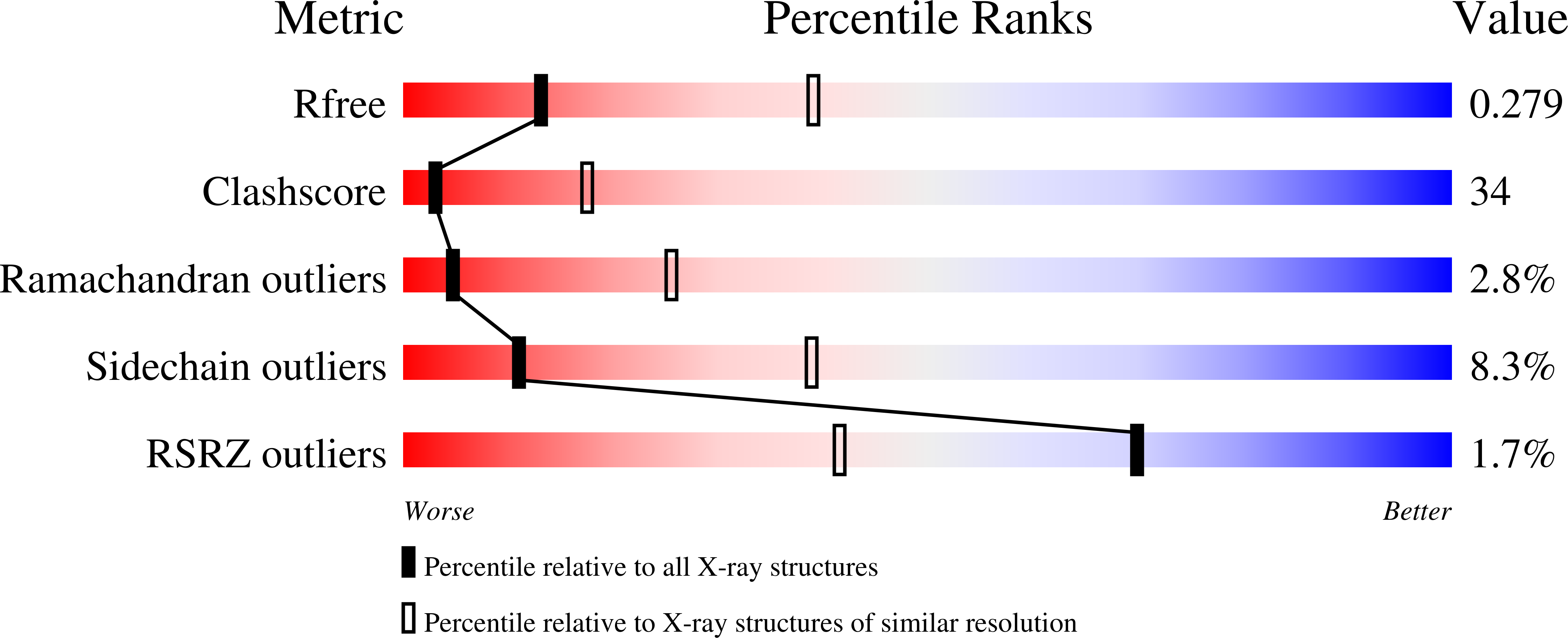
wwPDB Validation   3D Report Full Report


Ligand Structure Quality Assessment 


This is version 2.1 of the entry. See complete history


Literature

The Crystal Structure of the Quorum Sensing Protein Trar Bound to its Autoinducer and Target DNA

Vannini, A.Volpari, C.Gargioli, C.Muraglia, E.Cortese, R.De Francesco, R.Neddermann, P.Di Marco, S.

(2002) EMBO J 21: 4393

  • DOI: https://doi.org/10.1093/emboj/cdf459
  • Primary Citation of Related Structures:  
    1H0M

  • PubMed Abstract: 

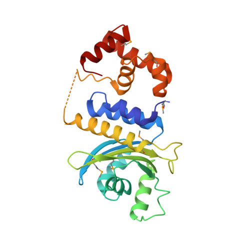
    The quorum sensing system allows bacteria to sense their cell density and initiate an altered pattern of gene expression after a sufficient quorum of cells has accumulated. In Agrobacterium tumefaciens, quorum sensing controls conjugal transfer of the tumour- inducing plasmid, responsible for plant crown gall disease. The core components of this system are the transcriptional regulator TraR and its inducing ligand N-(3-oxo-octanoyl)-L-homoserine lactone. This complex binds DNA and activates gene expression. We have determined the crystal structure of TraR in complex with its autoinducer and target DNA (PDB code 1h0m). The protein is dimeric, with each monomer composed of an N-terminal domain, which binds the ligand in an enclosed cavity far from the dimerization region, and a C-terminal domain, which binds DNA via a helix-turn-helix motif. The structure reveals an asymmetric homodimer, with one monomer longer than the other. The N-terminal domain resembles GAF/PAS domains, normally fused to catalytic signalling domains. In TraR, the gene fusion is between a GAF/PAS domain and a DNA-binding domain, resulting in a specific transcriptional regulator involved in quorum sensing.


  • Organizational Affiliation

    IRBM P. Angeletti, Via Pontina Km 30.600, I-00040 Pomezia, Rome, Italy.


Macromolecules

Find similar proteins by:  (by identity cutoff)  |  3D Structure
Entity ID: 1
MoleculeChains Sequence LengthOrganismDetailsImage
Transcriptional activator protein TraR
A, B, C, D
234Agrobacterium tumefaciensMutation(s): 0 
Gene Names: traR
UniProt
Find proteins for P33905 (Rhizobium radiobacter)
Explore P33905 
Go to UniProtKB:  P33905
Entity Groups  
Sequence Clusters30% Identity50% Identity70% Identity90% Identity95% Identity100% Identity
UniProt GroupP33905
Sequence Annotations
Expand
  • Reference Sequence

Find similar nucleic acids by:  Sequence   |   3D Structure  

Entity ID: 2
MoleculeChains LengthOrganismImage
5'-D(*AP*TP*GP*TP*GP*CP*AP*GP*AP*TP *CP*TP*GP*CP*AP*CP*AP*T)-3'
E, F, G, H
18synthetic construct
Sequence Annotations
Expand
  • Reference Sequence
Experimental Data & Validation

Experimental Data

  • Method: X-RAY DIFFRACTION
  • Resolution: 3.00 Å
  • R-Value Free: 0.285 
  • R-Value Work: 0.233 
  • Space Group: P 21 21 21
Unit Cell:
Length ( Å )Angle ( ˚ )
a = 66.993α = 90
b = 94.678β = 90
c = 209.669γ = 90
Software Package:
Software NamePurpose
MOSFLMdata reduction
SCALAdata scaling
SnBphasing
SHARPphasing
CNXrefinement

Structure Validation

View Full Validation Report


Ligand Structure Quality Assessment 


Entry History 

Deposition Data

Revision History  (Full details and data files)

  • Version 1.0: 2002-08-29
    Type: Initial release
  • Version 1.1: 2011-05-08
    Changes: Version format compliance
  • Version 1.2: 2011-07-13
    Changes: Version format compliance
  • Version 1.3: 2017-07-05
    Changes: Refinement description
  • Version 2.0: 2020-12-16
    Changes: Atomic model, Data collection, Database references, Derived calculations, Non-polymer description, Other, Source and taxonomy, Structure summary
  • Version 2.1: 2024-11-13
    Changes: Data collection, Database references, Structure summary