1H8D

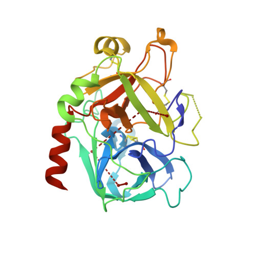
X-ray structure of the human alpha-thrombin complex with a tripeptide phosphonate inhibitor.


Experimental Data Snapshot

  • Method: X-RAY DIFFRACTION
  • Resolution: 1.40 Å
  • R-Value Free: 0.220 
  • R-Value Work: 0.170 

Starting Model: experimental
View more details

wwPDB Validation   3D Report Full Report


Ligand Structure Quality Assessment 


This is version 1.7 of the entry. See complete history


Literature

Inhibition of Human Alpha-Thrombin by a Phosphonate Tripeptide Proceeds Via a Metastable Pentacoordinated Phosphorus Intermediate

Skordalakes, E.Dodson, G.G.Green, D.S.Goodwin, C.A.Scully, M.F.Hudson, H.R.Kakkar, V.V.Deadman, J.J.

(2001) J Mol Biol 311: 549

  • DOI: https://doi.org/10.1006/jmbi.2001.4872
  • Primary Citation of Related Structures:  
    1H8D, 1H8I

  • PubMed Abstract: 

    X-ray crystallographic studies of human alpha-thrombin with a novel synthetic inhibitor, an acyl (alpha-aminoalkyl)phosphonate, reveal the existence of a pentacovalent phosphorus intermediate state. Crystal structures of the complex of alpha-thrombin with the phosphonate compound were determined independently using crystals of different ages. The first structure, solved from a crystal less than seven days old, showed a pentacoordinated phosphorus moiety. The second structure, determined from a crystal that was 12 weeks old, showed a tetracoordinated phosphorus moiety. In the first structure, a water molecule, made nucleophilic by coordination to His57 of alpha-thrombin, is bonded to the pentacoordinated phosphorus atom. Its position is approximately equivalent to that occupied by the water molecule responsible for hydrolytic deacylation during normal hydrolysis. The pentacoordinated phosphorus adduct collapses to give the expected pseudo tetrahedral complex, where the phosphorus atom is covalently bonded to Ser195 O(gamma). The crystallographic data presented here therefore suggest that the covalent bond formed between the inhibitor's phosphorus atom and O(gamma) of Ser195 proceeds via an addition-elimination mechanism, which involves the formation of a pentacoordinate intermediate.


  • Organizational Affiliation

    Chemistry Department and Biochemistry Department, Thrombosis Research Institute, Emmanuel Kaye Building, London, SW3 6LR, UK.


Macromolecules
Find similar proteins by:  (by identity cutoff)  |  3D Structure
Entity ID: 1
MoleculeChains Sequence LengthOrganismDetailsImage
THROMBINA [auth H]260Homo sapiensMutation(s): 0 
EC: 3.4.21.5
UniProt & NIH Common Fund Data Resources
Find proteins for P00734 (Homo sapiens)
Explore P00734 
Go to UniProtKB:  P00734
PHAROS:  P00734
GTEx:  ENSG00000180210 
Entity Groups  
Sequence Clusters30% Identity50% Identity70% Identity90% Identity95% Identity100% Identity
UniProt GroupP00734
Sequence Annotations
Expand
  • Reference Sequence

Find similar proteins by:  Sequence   |   3D Structure  

Entity ID: 2
MoleculeChains Sequence LengthOrganismDetailsImage
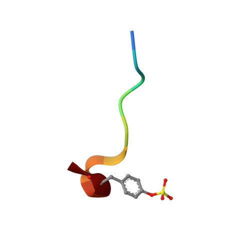
HIRUDIN IB [auth I]10Hirudo medicinalisMutation(s): 0 
UniProt
Find proteins for P01050 (Hirudo medicinalis)
Explore P01050 
Go to UniProtKB:  P01050
Entity Groups  
Sequence Clusters30% Identity50% Identity70% Identity90% Identity95% Identity100% Identity
UniProt GroupP01050
Sequence Annotations
Expand
  • Reference Sequence
Find similar proteins by:  (by identity cutoff)  |  3D Structure
Entity ID: 3
MoleculeChains Sequence LengthOrganismDetailsImage
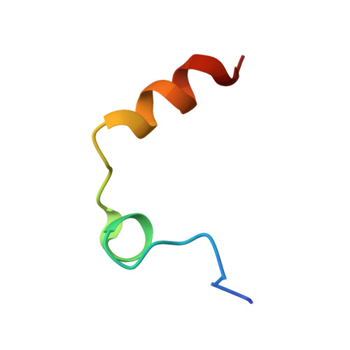
THROMBINC [auth L]29Homo sapiensMutation(s): 0 
EC: 3.4.21.5
UniProt & NIH Common Fund Data Resources
Find proteins for P00734 (Homo sapiens)
Explore P00734 
Go to UniProtKB:  P00734
PHAROS:  P00734
GTEx:  ENSG00000180210 
Entity Groups  
Sequence Clusters30% Identity50% Identity70% Identity90% Identity95% Identity100% Identity
UniProt GroupP00734
Sequence Annotations
Expand
  • Reference Sequence
Small Molecules
Ligands 1 Unique
IDChains Name / Formula / InChI Key2D Diagram3D Interactions
PHW
Query on PHW

Download Ideal Coordinates CCD File 
D [auth H]N-[(benzyloxy)carbonyl]-beta-phenyl-D-phenylalanyl-N-{(1S,3E)-1-[dihydroxy(diphenoxy)-lambda~5~-phosphanyl]-4-methoxybut-3-en-1-yl}-L-prolinamide
C45 H48 N3 O9 P
RYHYSOICLVLDDN-JBHUJMBHSA-N
Modified Residues  1 Unique
IDChains TypeFormula2D DiagramParent
TYS
Query on TYS
B [auth I]L-PEPTIDE LINKINGC9 H11 N O6 STYR
Experimental Data & Validation

Experimental Data

  • Method: X-RAY DIFFRACTION
  • Resolution: 1.40 Å
  • R-Value Free: 0.220 
  • R-Value Work: 0.170 
  • Space Group: C 1 2 1
Unit Cell:
Length ( Å )Angle ( ˚ )
a = 70.64α = 90
b = 71.6β = 100.35
c = 71.9γ = 90
Software Package:
Software NamePurpose
REFMACrefinement
DENZOdata reduction
SCALEPACKdata scaling
AMoREphasing

Structure Validation

View Full Validation Report


Ligand Structure Quality Assessment 


Entry History 

Deposition Data

Revision History  (Full details and data files)

  • Version 1.0: 2001-02-26
    Type: Initial release
  • Version 1.1: 2011-07-13
    Changes: Atomic model, Database references, Derived calculations, Non-polymer description, Structure summary, Version format compliance
  • Version 1.2: 2012-11-30
    Changes: Other
  • Version 1.3: 2013-03-13
    Changes: Other
  • Version 1.4: 2017-07-12
    Changes: Advisory, Derived calculations
  • Version 1.5: 2018-11-21
    Changes: Advisory, Data collection, Derived calculations
  • Version 1.6: 2023-12-13
    Changes: Data collection, Database references, Derived calculations, Other, Refinement description
  • Version 1.7: 2024-11-13
    Changes: Structure summary