1HXD

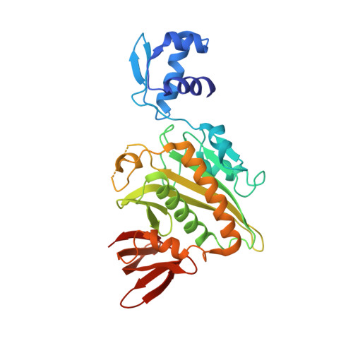
CRYSTAL STRUCTURE OF E. COLI BIOTIN REPRESSOR WITH BOUND BIOTIN


Experimental Data Snapshot

  • Method: X-RAY DIFFRACTION
  • Resolution: 2.40 Å
  • R-Value Work: 0.189 

Starting Model: experimental
View more details

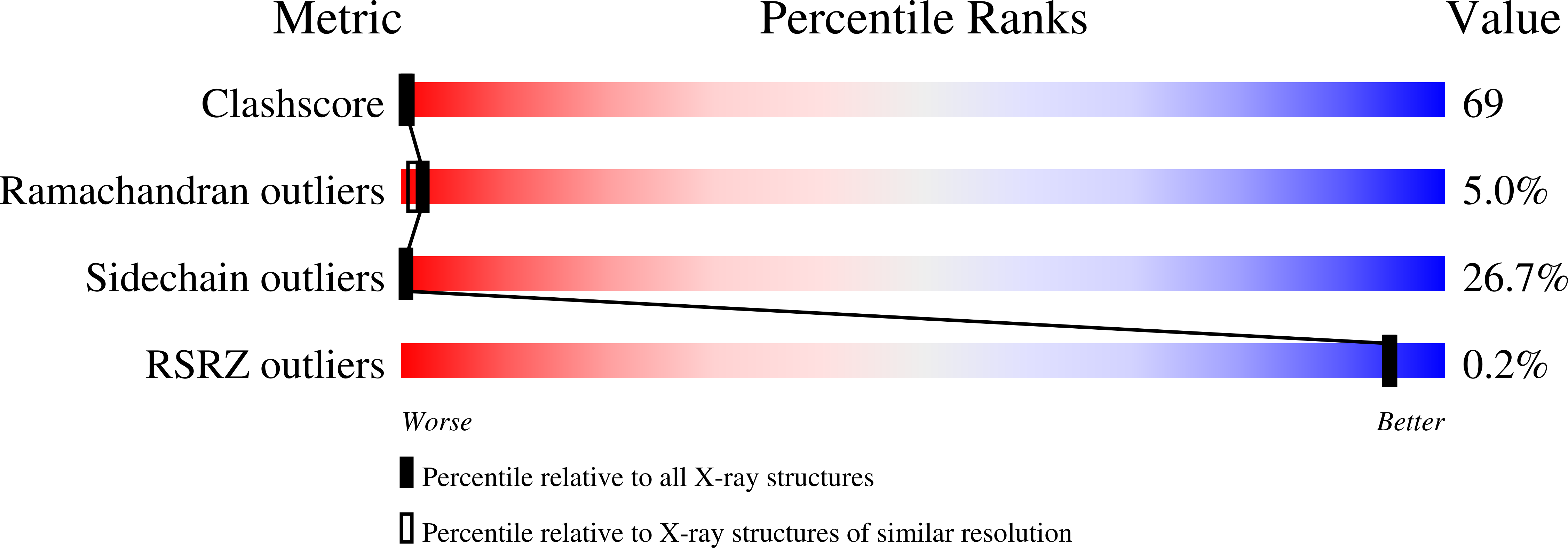
wwPDB Validation   3D Report Full Report


Ligand Structure Quality Assessment 


This is version 1.3 of the entry. See complete history


Literature

Corepressor-induced organization and assembly of the biotin repressor: a model for allosteric activation of a transcriptional regulator.

Weaver, L.H.Kwon, K.Beckett, D.Matthews, B.W.

(2001) Proc Natl Acad Sci U S A 98: 6045-6050

  • DOI: https://doi.org/10.1073/pnas.111128198
  • Primary Citation of Related Structures:  
    1HXD

  • PubMed Abstract: 

    The Escherichia coli biotin repressor binds to the biotin operator to repress transcription of the biotin biosynthetic operon. In this work, a structure determined by x-ray crystallography of a complex of the repressor bound to biotin, which also functions as an activator of DNA binding by the biotin repressor (BirA), is described. In contrast to the monomeric aporepressor, the complex is dimeric with an interface composed in part of an extended beta-sheet. Model building, coupled with biochemical data, suggests that this is the dimeric form of BirA that binds DNA. Segments of three surface loops that are disordered in the aporepressor structure are located in the interface region of the dimer and exhibit greater order than was observed in the aporepressor structure. The results suggest that the corepressor of BirA causes a disorder-to-order transition that is a prerequisite to repressor dimerization and DNA binding.


  • Organizational Affiliation

    Institute for Molecular Biology, Howard Hughes Medical Institute and Department of Physics, 1229 University of Oregon, Eugene, OR 97403-1229, USA. [email protected]


Macromolecules
Find similar proteins by:  (by identity cutoff)  |  3D Structure
Entity ID: 1
MoleculeChains Sequence LengthOrganismDetailsImage
BIRA BIFUNCTIONAL PROTEIN
A, B
321Escherichia coliMutation(s): 0 
EC: 6.3.4.15
UniProt
Find proteins for P06709 (Escherichia coli (strain K12))
Explore P06709 
Go to UniProtKB:  P06709
Entity Groups  
Sequence Clusters30% Identity50% Identity70% Identity90% Identity95% Identity100% Identity
UniProt GroupP06709
Sequence Annotations
Expand
  • Reference Sequence
Small Molecules
Binding Affinity Annotations 
IDSourceBinding Affinity
BTN BindingDB:  1HXD -TΔS: min: -6.68e+0, max: 40.95 (kJ/mol) from 6 assay(s)
ΔG: min: -5.81e+1, max: -4.10e+1 (kJ/mol) from 6 assay(s)
Experimental Data & Validation

Experimental Data

  • Method: X-RAY DIFFRACTION
  • Resolution: 2.40 Å
  • R-Value Work: 0.189 
  • Space Group: C 2 2 21
Unit Cell:
Length ( Å )Angle ( ˚ )
a = 104.9α = 90
b = 108.9β = 90
c = 143.2γ = 90
Software Package:
Software NamePurpose
TNTrefinement
SDMSdata reduction
SDMSdata scaling

Structure Validation

View Full Validation Report


Ligand Structure Quality Assessment 


Entry History 

Deposition Data

Revision History  (Full details and data files)

  • Version 1.0: 2001-05-30
    Type: Initial release
  • Version 1.1: 2008-04-27
    Changes: Version format compliance
  • Version 1.2: 2011-07-13
    Changes: Version format compliance
  • Version 1.3: 2023-08-09
    Changes: Data collection, Database references, Derived calculations, Refinement description