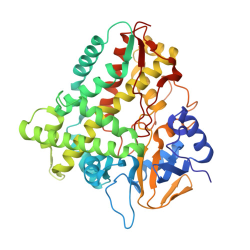
1JIP

P450eryF(A245S)/ketoconazole


Experimental Data Snapshot

  • Method: X-RAY DIFFRACTION
  • Resolution: 2.00 Å
  • R-Value Free: 0.247 
  • R-Value Work: 0.210 
  • R-Value Observed: 0.210 

Starting Model: experimental
View more details

wwPDB Validation   3D Report Full Report


Ligand Structure Quality Assessment 


This is version 1.5 of the entry. See complete history


Literature

Ketoconazole-induced conformational changes in the active site of cytochrome P450eryF.

Cupp-Vickery, J.R.Garcia, C.Hofacre, A.McGee-Estrada, K.

(2001) J Mol Biol 311: 101-110

  • DOI: https://doi.org/10.1006/jmbi.2001.4803
  • Primary Citation of Related Structures:  
    1JIN, 1JIO, 1JIP

  • PubMed Abstract: 

    The azole-based P450 inhibitor ketoconazole is used to treat fungal infections and functions by blocking ergosterol biosynthesis in yeast. Ketoconazole binds to mammalian P450 enzymes and this can result in drug-drug interactions and lead to liver damage. To identify protein-drug interactions that contribute to binding specificity and affinity, we determined the crystal structure of ketoconazole complexed with P450eryF. In the P450eryF/ketoconazole structure, the azole moiety and nearby rings of ketoconzole are positioned in the active site similar to the substrate, 6-deoxyerythronolide B, with the azole nitrogen atom coordinated to the heme iron atom. The remainder of the ketoconazole molecule extends into the active-site pocket, which is occupied by water in the substrate complex. Binding of ketoconazole led to unexpected conformational changes in the I-helix. The I-helix cleft near the active site has collapsed with a helical pitch of 5.4 A compared to 6.6 A in the substrate complex. P450eryF/ketoconazole crystals soaked in 6-deoxyerythronolide B to exchange ligands exhibit a structure identical with that of the original P450eryF/substrate complex, with the I-helix cleft restored to a pitch of 6.6 A. These findings indicate that the I-helix region of P450eryF is flexible and can adopt multiple conformations. An improved understanding of the flexibility of the active-site region of cytochrome P450 enzymes is important to gain insight into determinants of ligand binding/specificity as well as to evaluate models for catalytic mechanism based on static crystal structures.


  • Organizational Affiliation

    Department of Chemistry and Biochemistry, California State University, Fullerton, 800 N. State College Blvd., Fullerton, CA 92834, USA. [email protected]


Macromolecules
Find similar proteins by:  (by identity cutoff)  |  3D Structure
Entity ID: 1
MoleculeChains Sequence LengthOrganismDetailsImage
CYTOCHROME P450 107A1403Saccharopolyspora erythraeaMutation(s): 1 
EC: 1 (PDB Primary Data), 1.14.15.35 (UniProt)
UniProt
Find proteins for Q00441 (Saccharopolyspora erythraea (strain ATCC 11635 / DSM 40517 / JCM 4748 / NBRC 13426 / NCIMB 8594 / NRRL 2338))
Explore Q00441 
Go to UniProtKB:  Q00441
Entity Groups  
Sequence Clusters30% Identity50% Identity70% Identity90% Identity95% Identity100% Identity
UniProt GroupQ00441
Sequence Annotations
Expand
  • Reference Sequence
Small Molecules
Ligands 2 Unique
IDChains Name / Formula / InChI Key2D Diagram3D Interactions
HEM
Query on HEM

Download Ideal Coordinates CCD File 
B [auth A]PROTOPORPHYRIN IX CONTAINING FE
C34 H32 Fe N4 O4
KABFMIBPWCXCRK-RGGAHWMASA-L
KTN
Query on KTN

Download Ideal Coordinates CCD File 
C [auth A]CIS-1-ACETYL-4-(4-((2-(2,4-DICHLOROPHENYL)-2-(1H-IMIDAZOL-1-YLMETHYL)-1,3-DIOXOLAN-4-YL)METHOXY)PHENYL)PIPERAZINE
C26 H28 Cl2 N4 O4
XMAYWYJOQHXEEK-JYFHCDHNSA-N
Experimental Data & Validation

Experimental Data

  • Method: X-RAY DIFFRACTION
  • Resolution: 2.00 Å
  • R-Value Free: 0.247 
  • R-Value Work: 0.210 
  • R-Value Observed: 0.210 
  • Space Group: P 21 21 21
Unit Cell:
Length ( Å )Angle ( ˚ )
a = 54.23α = 90
b = 80.49β = 90
c = 99.72γ = 90
Software Package:
Software NamePurpose
CNSrefinement
MOSFLMdata reduction
CCP4data scaling
CNSphasing

Structure Validation

View Full Validation Report


Ligand Structure Quality Assessment 


Entry History 

Deposition Data

Revision History  (Full details and data files)

  • Version 1.0: 2001-10-17
    Type: Initial release
  • Version 1.1: 2008-04-27
    Changes: Version format compliance
  • Version 1.2: 2011-07-13
    Changes: Version format compliance
  • Version 1.3: 2021-10-27
    Changes: Database references, Derived calculations
  • Version 1.4: 2024-02-07
    Changes: Data collection
  • Version 1.5: 2024-04-03
    Changes: Refinement description