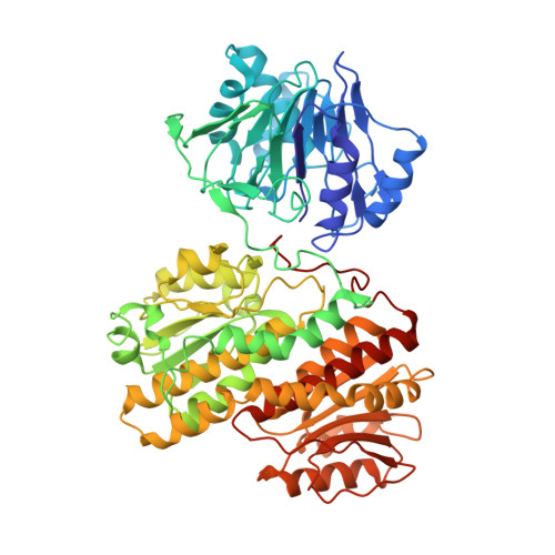
1JXA

GLUCOSAMINE 6-PHOSPHATE SYNTHASE WITH GLUCOSE 6-PHOSPHATE


Experimental Data Snapshot

  • Method: X-RAY DIFFRACTION
  • Resolution: 3.10 Å
  • R-Value Free: 0.280 
  • R-Value Work: 0.200 
  • R-Value Observed: 0.220 

Starting Models: experimental
View more details

wwPDB Validation   3D Report Full Report


Ligand Structure Quality Assessment 


This is version 1.4 of the entry. See complete history


Literature

Channeling of ammonia in glucosamine-6-phosphate synthase.

Teplyakov, A.Obmolova, G.Badet, B.Badet-Denisot, M.A.

(2001) J Mol Biol 313: 1093-1102

  • DOI: https://doi.org/10.1006/jmbi.2001.5094
  • Primary Citation of Related Structures:  
    1JXA

  • PubMed Abstract: 

    Glucosamine-6-phosphate synthase catalyses the first and rate-limiting step in hexosamine metabolism, converting fructose 6-phosphate into glucosamine 6-phosphate in the presence of glutamine. The crystal structure of the Escherichia coli enzyme reveals the domain organisation of the homodimeric molecule. The 18 A hydrophobic channel sequestered from the solvent connects the glutaminase and isomerase active sites, and provides a means of ammonia transfer from glutamine to sugar phosphate. The C-terminal decapeptide sandwiched between the two domains plays a central role in the transfer. Based on the structure, a mechanism of enzyme action and self-regulation is proposed. It involves large domain movements triggered by substrate binding that lead to the formation of the channel.


  • Organizational Affiliation

    European Molecular Biology Laboratory, Notkestr. 85, D-22603 Hamburg, Germany. [email protected]


Macromolecules
Find similar proteins by:  (by identity cutoff)  |  3D Structure
Entity ID: 1
MoleculeChains Sequence LengthOrganismDetailsImage
glucosamine 6-phosphate synthase
A, B, C
608Escherichia coliMutation(s): 0 
EC: 2.6.1.16
UniProt
Find proteins for P17169 (Escherichia coli (strain K12))
Explore P17169 
Go to UniProtKB:  P17169
Entity Groups  
Sequence Clusters30% Identity50% Identity70% Identity90% Identity95% Identity100% Identity
UniProt GroupP17169
Sequence Annotations
Expand
  • Reference Sequence
Experimental Data & Validation

Experimental Data

  • Method: X-RAY DIFFRACTION
  • Resolution: 3.10 Å
  • R-Value Free: 0.280 
  • R-Value Work: 0.200 
  • R-Value Observed: 0.220 
  • Space Group: C 1 2 1
Unit Cell:
Length ( Å )Angle ( ˚ )
a = 131.4α = 90
b = 112.4β = 96.4
c = 185.1γ = 90
Software Package:
Software NamePurpose
MOLREPphasing
REFMACrefinement
DENZOdata reduction
SCALEPACKdata scaling

Structure Validation

View Full Validation Report


Ligand Structure Quality Assessment 


Entry History 

Deposition Data

Revision History  (Full details and data files)

  • Version 1.0: 2001-11-21
    Type: Initial release
  • Version 1.1: 2008-04-27
    Changes: Version format compliance
  • Version 1.2: 2011-07-13
    Changes: Version format compliance
  • Version 1.3: 2018-10-03
    Changes: Data collection
  • Version 1.4: 2023-08-16
    Changes: Data collection, Database references, Derived calculations, Refinement description