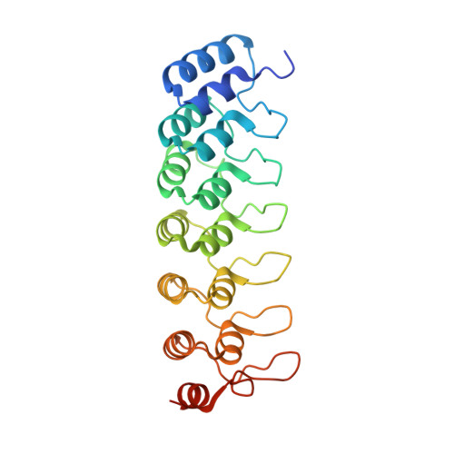
1K1A

Crystal structure of the ankyrin repeat domain of Bcl-3: a unique member of the IkappaB protein family


Experimental Data Snapshot

  • Method: X-RAY DIFFRACTION
  • Resolution: 1.86 Å
  • R-Value Free: 0.229 
  • R-Value Work: 0.199 
  • R-Value Observed: 0.200 

Starting Model: experimental
View more details

wwPDB Validation   3D Report Full Report


This is version 1.3 of the entry. See complete history


Literature

Crystal structure of the ankyrin repeat domain of Bcl-3: a unique member of the IkappaB protein family.

Michel, F.Soler-Lopez, M.Petosa, C.Cramer, P.Siebenlist, U.Muller, C.W.

(2001) EMBO J 20: 6180-6190

  • DOI: https://doi.org/10.1093/emboj/20.22.6180
  • Primary Citation of Related Structures:  
    1K1A, 1K1B

  • PubMed Abstract: 

    IkappaB proteins associate with the transcription factor NF-kappaB via their ankyrin repeat domain. Bcl-3 is an unusual IkappaB protein because it is primarily nucleoplasmic and can lead to enhanced NF-kappaB-dependent transcription, unlike the prototypical IkappaB protein IkappaBalpha, which inhibits NF-kappaB activity by retaining it in the cytoplasm. Here we report the 1.9 A crystal structure of the ankyrin repeat domain of human Bcl-3 and compare it with that of IkappaBalpha bound to NF-kappaB. The two structures are highly similar over the central ankyrin repeats but differ in the N-terminal repeat and at the C-terminus, where Bcl-3 contains a seventh repeat in place of the acidic PEST region of IkappaBalpha. Differences between the two structures suggest why Bcl-3 differs from IkappaBalpha in selectivity towards various NF-kappaB species, why Bcl-3 but not IkappaBalpha can associate with its NF-kappaB partner bound to DNA, and why two molecules of Bcl-3 but only one of IkappaBalpha can bind to its NF-kappaB partner. Comparison of the two structures thus provides an insight into the functional diversity of IkappaB proteins.


  • Organizational Affiliation

    European Molecular Biology Laboratory, Grenoble Outstation BP 181, 38042 Grenoble, Cedex 9, France.


Macromolecules
Find similar proteins by:  (by identity cutoff)  |  3D Structure
Entity ID: 1
MoleculeChains Sequence LengthOrganismDetailsImage
B-cell lymphoma 3-encoded protein241Homo sapiensMutation(s): 0 
UniProt & NIH Common Fund Data Resources
Find proteins for P20749 (Homo sapiens)
Explore P20749 
Go to UniProtKB:  P20749
PHAROS:  P20749
GTEx:  ENSG00000069399 
Entity Groups  
Sequence Clusters30% Identity50% Identity70% Identity90% Identity95% Identity100% Identity
UniProt GroupP20749
Sequence Annotations
Expand
  • Reference Sequence
Experimental Data & Validation

Experimental Data

  • Method: X-RAY DIFFRACTION
  • Resolution: 1.86 Å
  • R-Value Free: 0.229 
  • R-Value Work: 0.199 
  • R-Value Observed: 0.200 
  • Space Group: P 1 21 1
Unit Cell:
Length ( Å )Angle ( ˚ )
a = 31.703α = 90
b = 51.218β = 102.01
c = 64.68γ = 90
Software Package:
Software NamePurpose
AMoREphasing
REFMACrefinement
DENZOdata reduction
SCALEPACKdata scaling

Structure Validation

View Full Validation Report


Entry History 

Deposition Data

Revision History  (Full details and data files)

  • Version 1.0: 2001-11-21
    Type: Initial release
  • Version 1.1: 2008-04-27
    Changes: Version format compliance
  • Version 1.2: 2011-07-13
    Changes: Version format compliance
  • Version 1.3: 2023-08-16
    Changes: Data collection, Database references, Refinement description