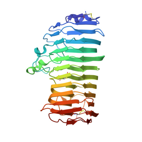
1KCD

Endopolygalacturonase I from Stereum purpureum complexed with two galacturonate at 1.15 A resolution.


Experimental Data Snapshot

  • Method: X-RAY DIFFRACTION
  • Resolution: 1.15 Å
  • R-Value Free: 0.164 
  • R-Value Work: 0.126 
  • R-Value Observed: 0.128 

Starting Model: experimental
View more details

wwPDB Validation   3D Report Full Report


Ligand Structure Quality Assessment 


This is version 3.1 of the entry. See complete history


Literature

Active-site architecture of endopolygalacturonase I from Stereum purpureum revealed by crystal structures in native and ligand-bound forms at atomic resolution.

Shimizu, T.Nakatsu, T.Miyairi, K.Okuno, T.Kato, H.

(2002) Biochemistry 41: 6651-6659

  • DOI: https://doi.org/10.1021/bi025541a
  • Primary Citation of Related Structures:  
    1K5C, 1KCC, 1KCD

  • PubMed Abstract: 

    Crystal structures of endopolygalacturonase from Stereum purpureum were solved in native and two galacturonic acid complex states at atomic resolution. Endopolygalacturonase catalyzes the hydrolysis of alpha-1,4-glycosidic linkage of polygalacturonate in pectin. The native structure was determined by the multiple wavelength anomalous dispersion method and was refined anisotropically with SHELXL-97, with an R factor of 11.4% and an R(free) factor of 14.0% at 0.96 A resolution. The enzyme folds into a right-handed parallel beta-helix with 10 complete turns. The crystal structures of its binary complex with one D-galacturonate and its ternary complex with two D-galacturonates were also determined to identify the substrate binding site at 1.0 and 1.15 A resolutions, respectively. In the binary complex, one beta-D-galactopyranuronate was found in the +1 subsite, thus proving the strong affinity of the +1 subsite expected from the bond cleavage frequency on oligogalacturonates. In the ternary complex, an additional beta-D-galactofuranuronate was found in the -1 subsite. In both subsites, the recognition of the galacturonate carboxy group is important in galacturonate binding. In the +1 subsite, the carboxy group interacts with three basic residues, His195, Arg226, and Lys228, which were conserved in all endopolygalacturonases. In the -1 subsite, the unique nonprolyl cis-peptide bond is believed to be involved in binding the carboxy group of the substrate. The active site architecture of the complexes provides insight into the mechanism of inverting glycosyl hydrolases and also sheds light on the basis of the differences between the family 28 and the other inverting glycosyl hydrolases.


  • Organizational Affiliation

    Kinetic Crystallography Research Team, Membrane Dynamics Research Group, RIKEN, Harima Institute at SPring-8, 1-1-1 Kouto, Mikazuki-cho, Sayo-gun, Hyogo 679-5148 Japan.


Macromolecules
Find similar proteins by:  (by identity cutoff)  |  3D Structure
Entity ID: 1
MoleculeChains Sequence LengthOrganismDetailsImage
ENDOPOLYGALACTURONASE335Chondrostereum purpureumMutation(s): 0 
EC: 3.2.1.15
UniProt
Find proteins for P79074 (Chondrostereum purpureum)
Explore P79074 
Go to UniProtKB:  P79074
Entity Groups  
Sequence Clusters30% Identity50% Identity70% Identity90% Identity95% Identity100% Identity
UniProt GroupP79074
Glycosylation
Glycosylation Sites: 2
Sequence Annotations
Expand
  • Reference Sequence
Small Molecules
Ligands 4 Unique
IDChains Name / Formula / InChI Key2D Diagram3D Interactions
NAG
Query on NAG

Download Ideal Coordinates CCD File 
D [auth A],
E [auth A]
2-acetamido-2-deoxy-beta-D-glucopyranose
C8 H15 N O6
OVRNDRQMDRJTHS-FMDGEEDCSA-N
GTR
Query on GTR

Download Ideal Coordinates CCD File 
C [auth A]beta-D-galactopyranuronic acid
C6 H10 O7
AEMOLEFTQBMNLQ-DTEWXJGMSA-N
GTK
Query on GTK

Download Ideal Coordinates CCD File 
B [auth A]beta-D-galactofuranuronic acid
C6 H10 O7
OEVWLAOZEALENB-ORELYVPDSA-N
CL
Query on CL

Download Ideal Coordinates CCD File 
F [auth A]CHLORIDE ION
Cl
VEXZGXHMUGYJMC-UHFFFAOYSA-M
Experimental Data & Validation

Experimental Data

  • Method: X-RAY DIFFRACTION
  • Resolution: 1.15 Å
  • R-Value Free: 0.164 
  • R-Value Work: 0.126 
  • R-Value Observed: 0.128 
  • Space Group: P 1
Unit Cell:
Length ( Å )Angle ( ˚ )
a = 46.5α = 72.39
b = 52.07β = 69.5
c = 37.23γ = 70.54
Software Package:
Software NamePurpose
MOSFLMdata reduction
SCALAdata scaling
REFMACrefinement
SHELXL-97refinement
CCP4data scaling

Structure Validation

View Full Validation Report


Ligand Structure Quality Assessment 


Entry History 

Deposition Data

Revision History  (Full details and data files)

  • Version 1.0: 2002-06-05
    Type: Initial release
  • Version 1.1: 2008-04-27
    Changes: Version format compliance
  • Version 1.2: 2011-07-13
    Changes: Non-polymer description, Version format compliance
  • Version 1.3: 2011-08-24
    Changes: Non-polymer description
  • Version 1.4: 2015-10-14
    Changes: Non-polymer description
  • Version 2.0: 2020-07-29
    Type: Remediation
    Reason: Carbohydrate remediation
    Changes: Atomic model, Data collection, Derived calculations, Structure summary
  • Version 2.1: 2023-10-25
    Changes: Data collection, Database references, Refinement description, Structure summary
  • Version 3.0: 2023-11-15
    Changes: Atomic model
  • Version 3.1: 2024-11-06
    Changes: Structure summary