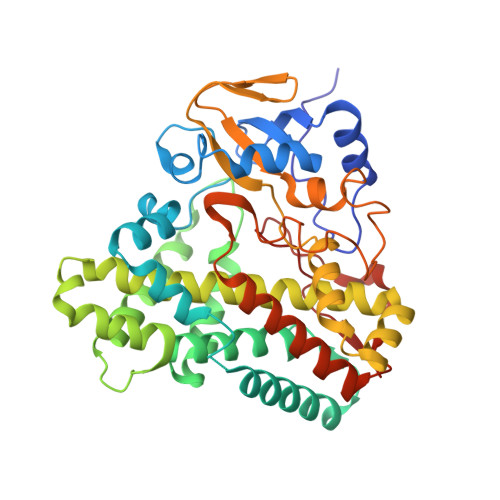
1N4G

Structure of CYP121, a Mycobacterial P450, in Complex with Iodopyrazole


Experimental Data Snapshot

  • Method: X-RAY DIFFRACTION
  • Resolution: 1.80 Å
  • R-Value Free: 0.217 
  • R-Value Work: 0.205 
  • R-Value Observed: 0.206 

Starting Model: experimental
View more details

wwPDB Validation   3D Report Full Report


Ligand Structure Quality Assessment 


This is version 1.3 of the entry. See complete history


Literature

Atomic structure of Mycobacterium tuberculosis CYP121 to 1.06 A reveals novel features of cytochrome P450.

Leys, D.Mowat, C.G.McLean, K.J.Richmond, A.Chapman, S.K.Walkinshaw, M.D.Munro, A.W.

(2003) J Biol Chem 278: 5141-5147

  • DOI: https://doi.org/10.1074/jbc.M209928200
  • Primary Citation of Related Structures:  
    1N40, 1N4G

  • PubMed Abstract: 

    The first structure of a P450 to an atomic resolution of 1.06 A has been solved for CYP121 from Mycobacterium tuberculosis. A comparison with P450 EryF (CYP107A1) reveals a remarkable overall similarity in fold with major differences residing in active site structural elements. The high resolution obtained allows visualization of several unusual aspects. The heme cofactor is bound in two distinct conformations while being notably kinked in one pyrrole group due to close interaction with the proline residue (Pro(346)) immediately following the heme iron-ligating cysteine (Cys(345)). The active site is remarkably rigid in comparison with the remainder of the structure, notwithstanding the large cavity volume of 1350 A(3). The region immediately surrounding the distal water ligand is remarkable in several aspects. Unlike other bacterial P450s, the I helix shows no deformation, similar to mammalian CYP2C5. In addition, the positively charged Arg(386) is located immediately above the heme plane, dominating the local structure. Putative proton relay pathways from protein surface to heme (converging at Ser(279)) are identified. Most interestingly, the electron density indicates weak binding of a dioxygen molecule to the P450. This structure provides a basis for rational design of putative antimycobacterial agents.


  • Organizational Affiliation

    Department of Biochemistry, University of Leicester, The Adrian Building, University Road, Leicester LE1 7RH, United Kingdom. [email protected]


Macromolecules
Find similar proteins by:  (by identity cutoff)  |  3D Structure
Entity ID: 1
MoleculeChains Sequence LengthOrganismDetailsImage
Cytochrome P450 121396Mycobacterium tuberculosisMutation(s): 0 
Gene Names: CYP121 OR RV2276 OR MT2336 OR MTCY339.34C
EC: 1.14 (PDB Primary Data), 1.14.19.70 (UniProt)
UniProt
Find proteins for P9WPP7 (Mycobacterium tuberculosis (strain ATCC 25618 / H37Rv))
Explore P9WPP7 
Go to UniProtKB:  P9WPP7
Entity Groups  
Sequence Clusters30% Identity50% Identity70% Identity90% Identity95% Identity100% Identity
UniProt GroupP9WPP7
Sequence Annotations
Expand
  • Reference Sequence
Experimental Data & Validation

Experimental Data

  • Method: X-RAY DIFFRACTION
  • Resolution: 1.80 Å
  • R-Value Free: 0.217 
  • R-Value Work: 0.205 
  • R-Value Observed: 0.206 
  • Space Group: P 65 2 2
Unit Cell:
Length ( Å )Angle ( ˚ )
a = 78.717α = 90
b = 78.717β = 90
c = 268.24γ = 120
Software Package:
Software NamePurpose
HKL-2000data collection
SCALEPACKdata scaling
AMoREphasing
REFMACrefinement
HKL-2000data reduction

Structure Validation

View Full Validation Report


Ligand Structure Quality Assessment 


Entry History 

Revision History  (Full details and data files)

  • Version 1.0: 2003-02-04
    Type: Initial release
  • Version 1.1: 2008-04-28
    Changes: Version format compliance
  • Version 1.2: 2011-07-13
    Changes: Version format compliance
  • Version 1.3: 2024-02-14
    Changes: Data collection, Database references, Derived calculations, Refinement description