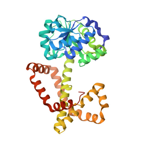
1NP3

Crystal structure of class I acetohydroxy acid isomeroreductase from Pseudomonas aeruginosa


Experimental Data Snapshot

  • Method: X-RAY DIFFRACTION
  • Resolution: 2.00 Å
  • R-Value Free: 0.235 
  • R-Value Work: 0.212 
  • R-Value Observed: 0.212 

wwPDB Validation   3D Report Full Report


This is version 1.3 of the entry. See complete history


Literature

Crystal Structure of Class I Acetohydroxy Acid Isomeroreductase from Pseudomonas aeruginosa

Ahn, H.J.Eom, S.J.Yoon, H.-J.Lee, B.I.Cho, H.Suh, S.W.

(2003) J Mol Biol 328: 505-515

  • DOI: https://doi.org/10.1016/s0022-2836(03)00264-x
  • Primary Citation of Related Structures:  
    1NP3

  • PubMed Abstract: 

    Acetohydroxy acid isomeroreductase (AHIR) is a key enzyme in the biosynthesis of branched-chain amino acids. We have determined the first crystal structure of a class I AHIR from Pseudomonas aeruginosa at 2.0 A resolution. Its dodecameric architecture of 23 point group symmetry is assembled of six dimeric units and dimerization is essential for the formation of the active site. The dimeric unit of P.aeruginosa AHIR partially superimposes with a three-domain monomer of spinach AHIR, a class II enzyme. This demonstrates that the so-called plant-specific insert in the middle of spinach AHIR is structurally and functionally equivalent to the C-terminal alpha-helical domain of P.aeruginosa AHIR, and the C-terminal alpha-helical domain was duplicated during evolution from the shorter, class I AHIRs to the longer, class II AHIRs. The dimeric unit of P.aeruginosa AHIR possesses a deep figure-of-eight knot, essentially identical with that in the spinach AHIR monomer. Thus, our work lowers the likelihood of the previous proposal that "domain duplication followed by exchange of a secondary structure element can be a source of such a knot in the protein structure" being correct.


  • Organizational Affiliation

    Structural Proteomics Laboratory, School of Chemistry and Molecular Engineering, College of Natural Sciences, Seoul National University, Seoul 151742, South Korea.


Macromolecules
Find similar proteins by:  (by identity cutoff)  |  3D Structure
Entity ID: 1
MoleculeChains Sequence LengthOrganismDetailsImage
Ketol-acid reductoisomerase
A, B, C, D
338Pseudomonas aeruginosaMutation(s): 0 
EC: 1.1.1.86
UniProt
Find proteins for Q9HVA2 (Pseudomonas aeruginosa (strain ATCC 15692 / DSM 22644 / CIP 104116 / JCM 14847 / LMG 12228 / 1C / PRS 101 / PAO1))
Explore Q9HVA2 
Go to UniProtKB:  Q9HVA2
Entity Groups  
Sequence Clusters30% Identity50% Identity70% Identity90% Identity95% Identity100% Identity
UniProt GroupQ9HVA2
Sequence Annotations
Expand
  • Reference Sequence
Experimental Data & Validation

Experimental Data

  • Method: X-RAY DIFFRACTION
  • Resolution: 2.00 Å
  • R-Value Free: 0.235 
  • R-Value Work: 0.212 
  • R-Value Observed: 0.212 
  • Space Group: P 21 3
Unit Cell:
Length ( Å )Angle ( ˚ )
a = 184.377α = 90
b = 184.377β = 90
c = 184.377γ = 90
Software Package:
Software NamePurpose
CNSrefinement

Structure Validation

View Full Validation Report


Entry History 

Deposition Data

Revision History  (Full details and data files)

  • Version 1.0: 2003-05-06
    Type: Initial release
  • Version 1.1: 2008-04-29
    Changes: Version format compliance
  • Version 1.2: 2011-07-13
    Changes: Derived calculations, Version format compliance
  • Version 1.3: 2024-03-13
    Changes: Data collection, Database references