1OEJ

YodA from Escherichia coli crystallised with no added ions


Experimental Data Snapshot

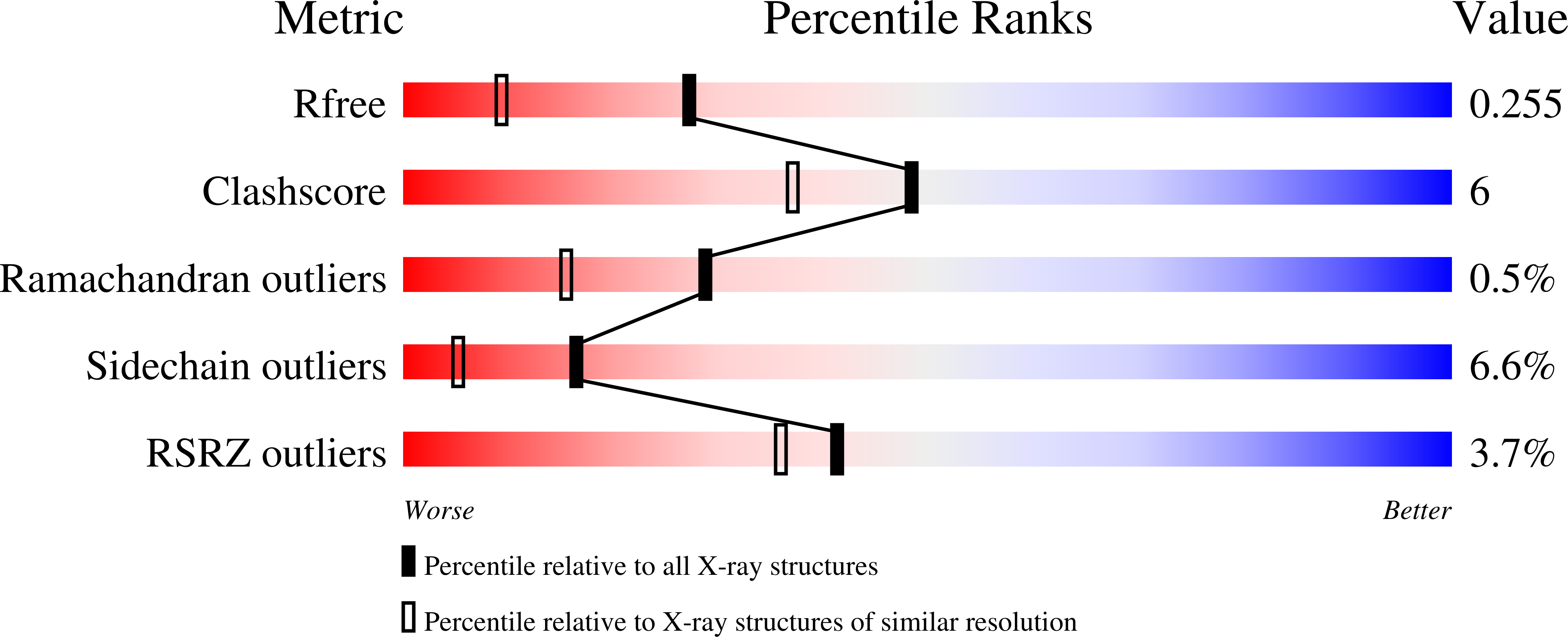
  • Method: X-RAY DIFFRACTION
  • Resolution: 1.81 Å
  • R-Value Free: 0.252 
  • R-Value Work: 0.180 
  • R-Value Observed: 0.184 

Starting Model: experimental
View more details

wwPDB Validation   3D Report Full Report


This is version 1.4 of the entry. See complete history


Literature

Yoda from Escherichia Coli is a Metal-Binding, Lipocalin-Like Protein

David, G.Blondeau, K.Schiltz, M.Penel, S.Lewit-Bentley, A.

(2003) J Biol Chem 278: 43728

  • DOI: https://doi.org/10.1074/jbc.M304484200
  • Primary Citation of Related Structures:  
    1OEE, 1OEJ, 1OEK

  • PubMed Abstract: 

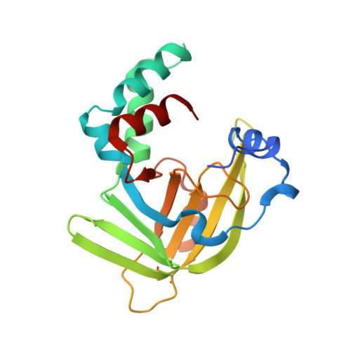
    We have determined the crystal structure of YodA, an Escherichia coli protein of unknown function. YodA had been identified under conditions of cadmium stress, and we confirm that it binds metals such as cadmium and zinc. We have also found nickel bound in one of the crystal forms. YodA is composed of two domains: a main lipocalin/calycin-like domain and a helical domain. The principal metal-binding site lies on one side of the calycin domain, thus making YodA the first metal-binding lipocalin known. Our experiments suggest that YodA expression may be part of a more general stress response. From sequence analogy with the C-terminal domain of a metal-binding receptor of a member of bacterial ATP-binding cassette transporters, we propose a three-dimensional model for this receptor and suggest that YodA may have a receptor-type partner in E. coli.


  • Organizational Affiliation

    Laboratoire pour l'Utilisation du Rayonnement Electromagnétique, CNRS, CEA, MdR, BP. 34, 91898 Orsay Cedex, France.


Macromolecules
Find similar proteins by:  (by identity cutoff)  |  3D Structure
Entity ID: 1
MoleculeChains Sequence LengthOrganismDetailsImage
HYPOTHETICAL PROTEIN YODA193Escherichia coliMutation(s): 0 
UniProt
Find proteins for P76344 (Escherichia coli (strain K12))
Explore P76344 
Go to UniProtKB:  P76344
Entity Groups  
Sequence Clusters30% Identity50% Identity70% Identity90% Identity95% Identity100% Identity
UniProt GroupP76344
Sequence Annotations
Expand
  • Reference Sequence
Small Molecules
Ligands 1 Unique
IDChains Name / Formula / InChI Key2D Diagram3D Interactions
NI
Query on NI

Download Ideal Coordinates CCD File 
B [auth A]NICKEL (II) ION
Ni
VEQPNABPJHWNSG-UHFFFAOYSA-N
Experimental Data & Validation

Experimental Data

  • Method: X-RAY DIFFRACTION
  • Resolution: 1.81 Å
  • R-Value Free: 0.252 
  • R-Value Work: 0.180 
  • R-Value Observed: 0.184 
  • Space Group: P 1 21 1
Unit Cell:
Length ( Å )Angle ( ˚ )
a = 40.348α = 90
b = 64.558β = 117.83
c = 41.499γ = 90
Software Package:
Software NamePurpose
MOSFLMdata reduction
SCALAdata scaling
AMoREphasing
CCP4phasing
REFMACrefinement

Structure Validation

View Full Validation Report


Entry History 

Deposition Data

Revision History  (Full details and data files)

  • Version 1.0: 2003-08-15
    Type: Initial release
  • Version 1.1: 2011-05-08
    Changes: Version format compliance
  • Version 1.2: 2011-07-13
    Changes: Version format compliance
  • Version 1.3: 2023-12-13
    Changes: Data collection, Database references, Derived calculations, Other, Refinement description
  • Version 1.4: 2024-11-06
    Changes: Structure summary