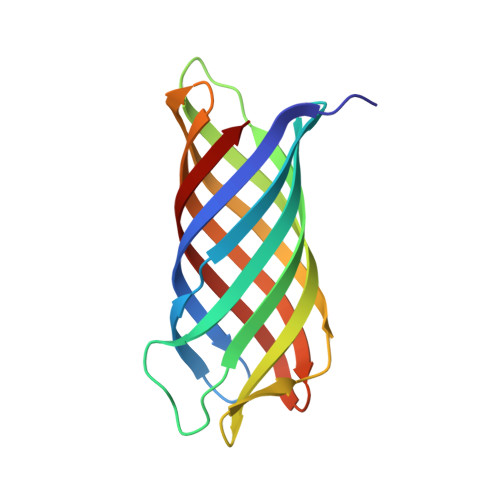
1P4T

Crystal structure of Neisserial surface protein A (NspA)


Experimental Data Snapshot

  • Method: X-RAY DIFFRACTION
  • Resolution: 2.55 Å
  • R-Value Free: 0.258 
  • R-Value Work: 0.225 
  • R-Value Observed: 0.227 

wwPDB Validation   3D Report Full Report


Ligand Structure Quality Assessment 


This is version 2.0 of the entry. See complete history


Literature

Crystal structure of Neisserial Surface Protein A (NspA), a conserved outer membrane protein with vaccine potential

Vandeputte-Rutten, L.Bos, M.P.Tommassen, J.Gros, P.

(2003) J Biol Chem 278: 24825-24830

  • DOI: https://doi.org/10.1074/jbc.M302803200
  • Primary Citation of Related Structures:  
    1P4T

  • PubMed Abstract: 

    The neisserial surface protein A (NspA) from Neisseria meningitidis is a promising vaccine candidate because it is highly conserved among meningococcal strains and induces bactericidal antibodies. NspA is a homolog of the Opa proteins, which mediate adhesion to host cells. Here, we present the crystal structure of NspA, determined to 2.55-A resolution. NspA forms an eight-stranded antiparallel beta-barrel. The four loops at the extracellular side of the NspA molecule form a long cleft, which contains mainly hydrophobic residues and harbors a detergent molecule, suggesting that the protein might function in the binding of hydrophobic ligands, such as lipids. In addition, the structure provides a starting point for structure-based vaccine design.


  • Organizational Affiliation

    Department of Crystal and Structural Chemistry, Bijvoet Center for Biomolecular Research, Utrecht, The Netherlands.


Macromolecules
Find similar proteins by:  (by identity cutoff)  |  3D Structure
Entity ID: 1
MoleculeChains Sequence LengthOrganismDetailsImage
outer membrane protein NspA155Neisseria meningitidisMutation(s): 0 
Gene Names: nspa
Membrane Entity: Yes 
UniProt
Find proteins for Q9RP17 (Neisseria meningitidis)
Explore Q9RP17 
Go to UniProtKB:  Q9RP17
Entity Groups  
Sequence Clusters30% Identity50% Identity70% Identity90% Identity95% Identity100% Identity
UniProt GroupQ9RP17
Sequence Annotations
Expand
  • Reference Sequence
Small Molecules
Ligands 3 Unique
IDChains Name / Formula / InChI Key2D Diagram3D Interactions
CXE
Query on CXE

Download Ideal Coordinates CCD File 
C [auth A],
D [auth A],
E [auth A],
F [auth A],
G [auth A]
PENTAETHYLENE GLYCOL MONODECYL ETHER
C20 H42 O6
QAXPOSPGRHYIHE-UHFFFAOYSA-N
SO4
Query on SO4

Download Ideal Coordinates CCD File 
B [auth A]SULFATE ION
O4 S
QAOWNCQODCNURD-UHFFFAOYSA-L
ETA
Query on ETA

Download Ideal Coordinates CCD File 
H [auth A]ETHANOLAMINE
C2 H7 N O
HZAXFHJVJLSVMW-UHFFFAOYSA-N
Experimental Data & Validation

Experimental Data

  • Method: X-RAY DIFFRACTION
  • Resolution: 2.55 Å
  • R-Value Free: 0.258 
  • R-Value Work: 0.225 
  • R-Value Observed: 0.227 
  • Space Group: H 3 2
Unit Cell:
Length ( Å )Angle ( ˚ )
a = 97.372α = 90
b = 97.372β = 90
c = 171.939γ = 120
Software Package:
Software NamePurpose
REFMACrefinement
DENZOdata reduction
SCALEPACKdata scaling
MLPHAREphasing

Structure Validation

View Full Validation Report


Ligand Structure Quality Assessment 


Entry History 

Deposition Data

Revision History  (Full details and data files)

  • Version 1.0: 2003-07-22
    Type: Initial release
  • Version 1.1: 2008-04-29
    Changes: Version format compliance
  • Version 1.2: 2011-07-13
    Changes: Advisory, Derived calculations, Version format compliance
  • Version 2.0: 2023-11-15
    Changes: Atomic model, Data collection, Database references, Derived calculations