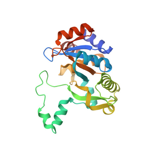
1PVY

3,4-dihydroxy-2-butanone 4-phosphate synthase from M. jannaschii in complex with ribulose 5-phosphate


Experimental Data Snapshot

  • Method: X-RAY DIFFRACTION
  • Resolution: 1.70 Å
  • R-Value Free: 0.259 
  • R-Value Work: 0.213 
  • R-Value Observed: 0.223 

Starting Model: experimental
View more details

wwPDB Validation   3D Report Full Report


Ligand Structure Quality Assessment 


This is version 1.4 of the entry. See complete history


Literature

Structure of 3,4-Dihydroxy-2-butanone 4-Phosphate Synthase from Methanococcus jannaschii in Complex with Divalent Metal Ions and the Substrate Ribulose 5-Phosphate: IMPLICATIONS FOR THE CATALYTIC MECHANISM

Steinbacher, S.Schiffmann, S.Richter, G.Huber, R.Bacher, A.Fischer, M.

(2003) J Biol Chem 278: 42256-42265

  • DOI: https://doi.org/10.1074/jbc.M307301200
  • Primary Citation of Related Structures:  
    1PVW, 1PVY

  • PubMed Abstract: 

    Skeletal rearrangements of carbohydrates are crucial for many biosynthetic pathways. In riboflavin biosynthesis ribulose 5-phosphate is converted into 3,4-dihydroxy-2-butanone 4-phosphate while its C4 atom is released as formate in a sequence of metal-dependent reactions. Here, we present the crystal structure of Methanococcus jannaschii 3,4-dihydroxy-2-butanone 4-phosphate synthase in complex with the substrate ribulose 5-phosphate at a dimetal center presumably consisting of non-catalytic zinc and calcium ions at 1.7-A resolution. The carbonyl group (O2) and two out of three free hydroxyl groups (OH3 and OH4) of the substrate are metal-coordinated. We correlate previous mutational studies on this enzyme with the present structural results. Residues of the first coordination sphere involved in metal binding are indispensable for catalytic activity. Only Glu-185 of the second coordination sphere cannot be replaced without complete loss of activity. It contacts the C3 hydrogen atom directly and probably initiates enediol formation in concert with both metal ions to start the reaction sequence. Mechanistic similarities to Rubisco acting on the similar substrate ribulose 1,5-diphosphate in carbon dioxide fixation as well as other carbohydrate (reducto-) isomerases are discussed.


  • Organizational Affiliation

    Max-Planck-Institut für Biochemie, Abteilung für Strukturforschung, Am Klopferspitz 18a, D-82152 Martinsried, Germany. [email protected]


Macromolecules
Find similar proteins by:  (by identity cutoff)  |  3D Structure
Entity ID: 1
MoleculeChains Sequence LengthOrganismDetailsImage
3,4-dihydroxy-2-butanone 4-phosphate synthase
A, B
227Methanocaldococcus jannaschiiMutation(s): 1 
Gene Names: MJ0055
EC: 5.4.99 (PDB Primary Data), 4.1.99.12 (UniProt)
UniProt
Find proteins for Q60364 (Methanocaldococcus jannaschii (strain ATCC 43067 / DSM 2661 / JAL-1 / JCM 10045 / NBRC 100440))
Explore Q60364 
Go to UniProtKB:  Q60364
Entity Groups  
Sequence Clusters30% Identity50% Identity70% Identity90% Identity95% Identity100% Identity
UniProt GroupQ60364
Sequence Annotations
Expand
  • Reference Sequence
Small Molecules
Ligands 3 Unique
IDChains Name / Formula / InChI Key2D Diagram3D Interactions
5RP
Query on 5RP

Download Ideal Coordinates CCD File 
C [auth A],
I [auth B]
RIBULOSE-5-PHOSPHATE
C5 H11 O8 P
FNZLKVNUWIIPSJ-UHNVWZDZSA-N
ZN
Query on ZN

Download Ideal Coordinates CCD File 
D [auth A],
H [auth A],
J [auth B]
ZINC ION
Zn
PTFCDOFLOPIGGS-UHFFFAOYSA-N
CA
Query on CA

Download Ideal Coordinates CCD File 
E [auth A],
F [auth A],
G [auth A],
K [auth B]
CALCIUM ION
Ca
BHPQYMZQTOCNFJ-UHFFFAOYSA-N
Experimental Data & Validation

Experimental Data

  • Method: X-RAY DIFFRACTION
  • Resolution: 1.70 Å
  • R-Value Free: 0.259 
  • R-Value Work: 0.213 
  • R-Value Observed: 0.223 
  • Space Group: P 1 21 1
Unit Cell:
Length ( Å )Angle ( ˚ )
a = 53.197α = 90
b = 69.272β = 93.52
c = 57.11γ = 90
Software Package:
Software NamePurpose
DENZOdata reduction
SCALEPACKdata scaling
AMoREphasing
CNSrefinement

Structure Validation

View Full Validation Report


Ligand Structure Quality Assessment 


Entry History 

Deposition Data

Revision History  (Full details and data files)

  • Version 1.0: 2003-11-04
    Type: Initial release
  • Version 1.1: 2008-04-29
    Changes: Version format compliance
  • Version 1.2: 2011-07-13
    Changes: Version format compliance
  • Version 1.3: 2021-11-10
    Changes: Database references, Derived calculations
  • Version 1.4: 2023-10-25
    Changes: Data collection, Refinement description