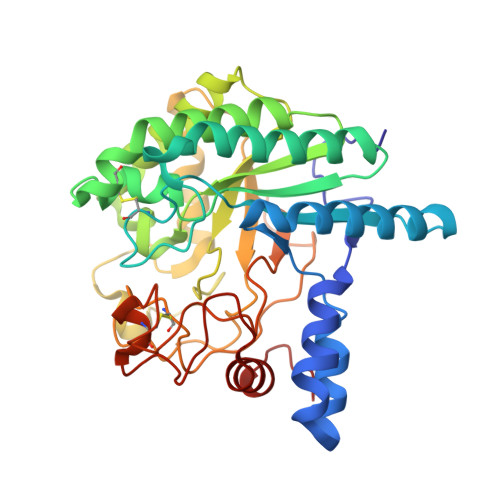
1QK2

WILD TYPE CEL6A WITH A NON-HYDROLYSABLE CELLOTETRAOSE


Experimental Data Snapshot

  • Method: X-RAY DIFFRACTION
  • Resolution: 2.00 Å
  • R-Value Free: 0.222 
  • R-Value Work: 0.189 
  • R-Value Observed: 0.189 

wwPDB Validation   3D Report Full Report


Ligand Structure Quality Assessment 


This is version 2.1 of the entry. See complete history


Literature

Crystallographic Evidence for Substrate Ring Distortion and Protein Conformational Changes During Catalysis in Cellobiohydrolase Cel6A from Trichoderma Reesei

Zou, J.-Y.Kleywegt, G.J.Stahlberg, J.Driguez, H.Nerinckx, W.Claeyssens, M.Koivula, A.Teeri, T.T.Jones, T.A.

(1999) Structure 7: 1035

  • DOI: https://doi.org/10.1016/s0969-2126(99)80171-3
  • Primary Citation of Related Structures:  
    1QJW, 1QK0, 1QK2

  • PubMed Abstract: 

    Cel6A is one of the two cellobiohydrolases produced by Trichoderma reesei. The catalytic core has a structure that is a variation of the classic TIM barrel. The active site is located inside a tunnel, the roof of which is formed mainly by a pair of loops. We describe three new ligand complexes. One is the structure of the wild-type enzyme in complex with a nonhydrolysable cello-oligosaccharide, methyl 4-S-beta-cellobiosyl-4-thio-beta-cellobioside (Glc)(2)-S-(Glc)(2), which differs from a cellotetraose in the nature of the central glycosidic linkage where a sulphur atom replaces an oxygen atom. The second structure is a mutant, Y169F, in complex with the same ligand, and the third is the wild-type enzyme in complex with m-iodobenzyl beta-D-glucopyranosyl-beta(1,4)-D-xylopyranoside (IBXG). The (Glc)(2)-S-(Glc)(2) ligand binds in the -2 to +2 sites in both the wild-type and mutant enzymes. The glucosyl unit in the -1 site is distorted from the usual chair conformation in both structures. The IBXG ligand binds in the -2 to +1 sites, with the xylosyl unit in the -1 site where it adopts the energetically favourable chair conformation. The -1 site glucosyl of the (Glc)(2)-S-(Glc)(2) ligand is unable to take on this conformation because of steric clashes with the protein. The crystallographic results show that one of the tunnel-forming loops in Cel6A is sensitive to modifications at the active site, and is able to take on a number of different conformations. One of the conformational changes disrupts a set of interactions at the active site that we propose is an integral part of the reaction mechanism.


  • Organizational Affiliation

    Department of Cell and Molecular Biology Uppsala University BMC Box 596, S-751 24, Uppsala, Sweden.


Macromolecules
Find similar proteins by:  (by identity cutoff)  |  3D Structure
Entity ID: 1
MoleculeChains Sequence LengthOrganismDetailsImage
CELLOBIOHYDROLASE CEL6A (FORMERLY CALLED CBH II)
A, B
363Trichoderma reeseiMutation(s): 0 
Gene Names: CBH2
EC: 3.2.1.91
UniProt
Find proteins for P07987 (Hypocrea jecorina)
Explore P07987 
Go to UniProtKB:  P07987
Entity Groups  
Sequence Clusters30% Identity50% Identity70% Identity90% Identity95% Identity100% Identity
UniProt GroupP07987
Glycosylation
Glycosylation Sites: 9
Sequence Annotations
Expand
  • Reference Sequence
Oligosaccharides

Help

Entity ID: 2
MoleculeChains Length2D Diagram Glycosylation3D Interactions
alpha-D-glucopyranose-(1-4)-alpha-D-glucopyranose-(1-4)-4-thio-beta-D-glucopyranose-(1-4)-methyl beta-D-glucopyranoside
C, D
4N/AN/A
Glycosylation Resources
GlyTouCan:  G73415XL
GlyCosmos:  G73415XL
Small Molecules
Ligands 2 Unique
IDChains Name / Formula / InChI Key2D Diagram3D Interactions
NAG
Query on NAG

Download Ideal Coordinates CCD File 
E [auth A],
F [auth A],
N [auth B],
O [auth B]
2-acetamido-2-deoxy-beta-D-glucopyranose
C8 H15 N O6
OVRNDRQMDRJTHS-FMDGEEDCSA-N
MAN
Query on MAN

Download Ideal Coordinates CCD File 
G [auth A]
H [auth A]
I [auth A]
J [auth A]
K [auth A]
G [auth A],
H [auth A],
I [auth A],
J [auth A],
K [auth A],
L [auth A],
M [auth A],
P [auth B],
Q [auth B],
R [auth B],
S [auth B],
T [auth B],
U [auth B],
V [auth B]
alpha-D-mannopyranose
C6 H12 O6
WQZGKKKJIJFFOK-PQMKYFCFSA-N
Experimental Data & Validation

Experimental Data

  • Method: X-RAY DIFFRACTION
  • Resolution: 2.00 Å
  • R-Value Free: 0.222 
  • R-Value Work: 0.189 
  • R-Value Observed: 0.189 
  • Space Group: P 1 21 1
Unit Cell:
Length ( Å )Angle ( ˚ )
a = 48.83α = 90
b = 74.7β = 104.12
c = 91.84γ = 90
Software Package:
Software NamePurpose
CNSrefinement
DENZOdata reduction
SCALEPACKdata scaling
CNSphasing

Structure Validation

View Full Validation Report


Ligand Structure Quality Assessment 


Entry History 

Deposition Data

Revision History  (Full details and data files)

  • Version 1.0: 1999-09-18
    Type: Initial release
  • Version 1.1: 2016-01-27
    Changes: Data collection, Database references, Derived calculations, Non-polymer description, Other, Refinement description, Source and taxonomy, Version format compliance
  • Version 1.2: 2019-10-23
    Changes: Author supporting evidence, Data collection, Derived calculations, Experimental preparation, Other, Structure summary
  • Version 2.0: 2020-07-29
    Type: Remediation
    Reason: Carbohydrate remediation
    Changes: Advisory, Atomic model, Data collection, Derived calculations, Structure summary
  • Version 2.1: 2024-10-23
    Changes: Data collection, Database references, Derived calculations, Structure summary