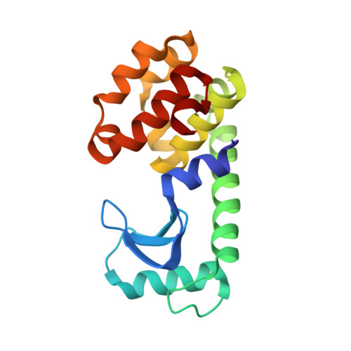
The introduction of strain and its effects on the structure and stability of T4 lysozyme.
Liu, R., Baase, W.A., Matthews, B.W.(2000) J Mol Biol 295: 127-145
- PubMed: 10623513
- DOI: https://doi.org/10.1006/jmbi.1999.3300
- Primary Citation of Related Structures:
1QS5, 1QS9, 1QSB, 1QTB, 1QTC, 1QTD, 1QTH - PubMed Abstract:
In order to try to better understand the role played by strain in the structure and stability of a protein a series of "small-to-large" mutations was made within the core of T4 lysozyme. Three different alanine residues, one involved in backbone contacts, one in side-chain contacts, and the third adjacent to a small cavity, were each replaced with subsets of the larger residues, Val, Leu, Ile, Met, Phe and Trp. As expected, the protein is progressively destabilized as the size of the introduced side-chain becomes larger. There does, however, seem to be a limit to the destabilization, suggesting that a protein of a given size may be capable of maintaining only a certain amount of strain. The changes in stability vary greatly from site to site. Substitution of larger residues for both Ala42 and Ala98 substantially destabilize the protein, even though the primary contacts in one case are predominantly with side-chain atoms and in the other with backbone. The results suggest that it is neither practical nor meaningful to try to separate the effects of introduced strain on side-chains from the effects on the backbone. Substitutions at Ala129 are much less destabilizing than at sites 42 or 98. This is most easily understood in terms of the pre-existing cavity, which provides partial space to accommodate the introduced side-chains. Crystal structures were obtained for a number of the mutants. These show that the changes in structure to accommodate the introduced side-chains usually consist of essentially rigid-body displacements of groups of linked atoms, achieved through relatively small changes in torsion angles. On rare occasions, a side-chain close to the site of substitution may change to a different rotamer. When such rotomer changes occur, they permit the structure to dissipate strain by a response that is plastic rather than elastic. In one case, a surface loop moves 1.2 A, not in direct response to a mutation, but in an interaction mediated via an intermolecular contact. It illustrates how the structure of a protein can be modified by crystal contacts.
Organizational Affiliation:
Institute of Molecular Biology, Howard Hughes Medical Institute and Department of Physics, Eugene, OR, 97403, USA.


