1R6D

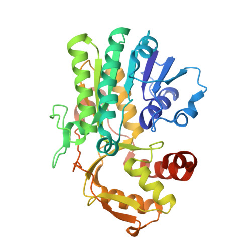
Crystal Structure of DesIV double mutant (dTDP-glucose 4,6-dehydratase) from Streptomyces venezuelae with NAD and DAU bound


Experimental Data Snapshot

  • Method: X-RAY DIFFRACTION
  • Resolution: 1.35 Å
  • R-Value Free: 0.216 
  • R-Value Work: 0.174 
  • R-Value Observed: 0.176 

Starting Model: experimental
View more details

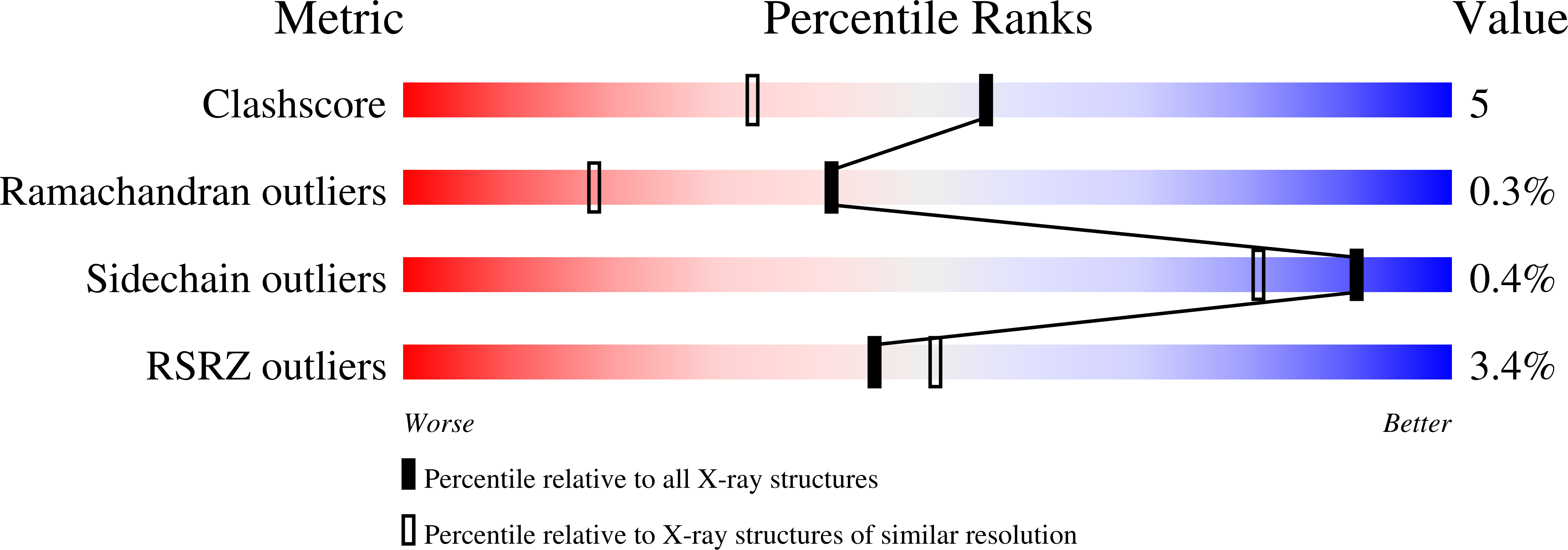
wwPDB Validation   3D Report Full Report


Ligand Structure Quality Assessment 


This is version 1.6 of the entry. See complete history


Literature

High Resolution X-ray Structure of dTDP-Glucose 4,6-Dehydratase from Streptomyces venezuelae

Allard, S.T.M.Cleland, W.W.Holden, H.M.

(2004) J Biol Chem 279: 2211-2220

  • DOI: https://doi.org/10.1074/jbc.M310134200
  • Primary Citation of Related Structures:  
    1R66, 1R6D

  • PubMed Abstract: 

    Desosamine is a 3-(dimethylamino)-3,4,6-trideoxyhexose found in some macrolide antibiotics. In Streptomyces venezuelae, there are seven genes required for the biosynthesis of this unusual sugar. One of the genes, desIV, codes for a dTDP-glucose 4,6-dehydratase, which is referred to as DesIV. The reaction mechanisms for these types of dehydratases are quite complicated with proton abstraction from the sugar 4'-hydroxyl group and hydride transfer to NAD+, proton abstraction at C-5, and elimination of the hydroxyl group at C-6 of the sugar, and finally return of a proton to C-5 and a hydride from NADH to C-6. Here we describe the cloning, overexpression, and purification, and high resolution x-ray crystallographic analysis to 1.44 A of wild-type DesIV complexed with dTDP. Additionally, for this study, a double site-directed mutant protein (D128N/E129Q) was prepared, crystallized as a complex with NAD+ and the substrate dTDP-glucose and its structure determined to 1.35 A resolution. In DesIV, the phenolate group of Tyr(151) and O(gamma) of Thr(127) lie at 2.7 and 2.6 A, respectively from the 4'-hydroxyl group of the dTDP-glucose substrate. The side chain of Asp(128) is in the correct position to function as a general acid for proton donation to the 6'-hydroxyl group while the side chain of Glu(129) is ideally situated to serve as the general base for proton abstraction at C-5. This investigation provides further detailed information for understanding the exquisite chemistry that occurs in these remarkable enzymes.


  • Organizational Affiliation

    Department of Biochemistry, University of Wisconsin, Madison, WI 53706, USA.


Macromolecules
Find similar proteins by:  (by identity cutoff)  |  3D Structure
Entity ID: 1
MoleculeChains Sequence LengthOrganismDetailsImage
TDP-glucose-4,6-dehydratase337Streptomyces venezuelaeMutation(s): 3 
Gene Names: DesIV
EC: 4.2.1.46
UniProt
Find proteins for Q9ZGH3 (Streptomyces venezuelae)
Explore Q9ZGH3 
Go to UniProtKB:  Q9ZGH3
Entity Groups  
Sequence Clusters30% Identity50% Identity70% Identity90% Identity95% Identity100% Identity
UniProt GroupQ9ZGH3
Sequence Annotations
Expand
  • Reference Sequence
Small Molecules
Ligands 2 Unique
IDChains Name / Formula / InChI Key2D Diagram3D Interactions
NAD
Query on NAD

Download Ideal Coordinates CCD File 
B [auth A]NICOTINAMIDE-ADENINE-DINUCLEOTIDE
C21 H27 N7 O14 P2
BAWFJGJZGIEFAR-NNYOXOHSSA-N
DAU
Query on DAU

Download Ideal Coordinates CCD File 
C [auth A]2'DEOXY-THYMIDINE-5'-DIPHOSPHO-ALPHA-D-GLUCOSE
C16 H26 N2 O16 P2
YSYKRGRSMLTJNL-URARBOGNSA-N
Experimental Data & Validation

Experimental Data

  • Method: X-RAY DIFFRACTION
  • Resolution: 1.35 Å
  • R-Value Free: 0.216 
  • R-Value Work: 0.174 
  • R-Value Observed: 0.176 
  • Space Group: P 21 21 2
Unit Cell:
Length ( Å )Angle ( ˚ )
a = 71.7α = 90
b = 99.4β = 90
c = 42.2γ = 90
Software Package:
Software NamePurpose
FRAMBOdata collection
XSCALIBREdata reduction
AMoREphasing
TNTrefinement
XSCALIBREdata scaling

Structure Validation

View Full Validation Report


Ligand Structure Quality Assessment 


Entry History 

Deposition Data

Revision History  (Full details and data files)

  • Version 1.0: 2004-01-27
    Type: Initial release
  • Version 1.1: 2008-04-29
    Changes: Version format compliance
  • Version 1.2: 2011-07-13
    Changes: Version format compliance
  • Version 1.3: 2012-05-23
    Changes: Atomic model
  • Version 1.4: 2017-10-11
    Changes: Refinement description
  • Version 1.5: 2021-10-27
    Changes: Database references, Derived calculations
  • Version 1.6: 2023-08-23
    Changes: Data collection, Refinement description