1RI2

Structure and mechanism of mRNA cap (guanine N-7) methyltransferase


Experimental Data Snapshot

  • Method: X-RAY DIFFRACTION
  • Resolution: 2.70 Å
  • R-Value Free: 0.295 
  • R-Value Work: 0.224 
  • R-Value Observed: 0.224 

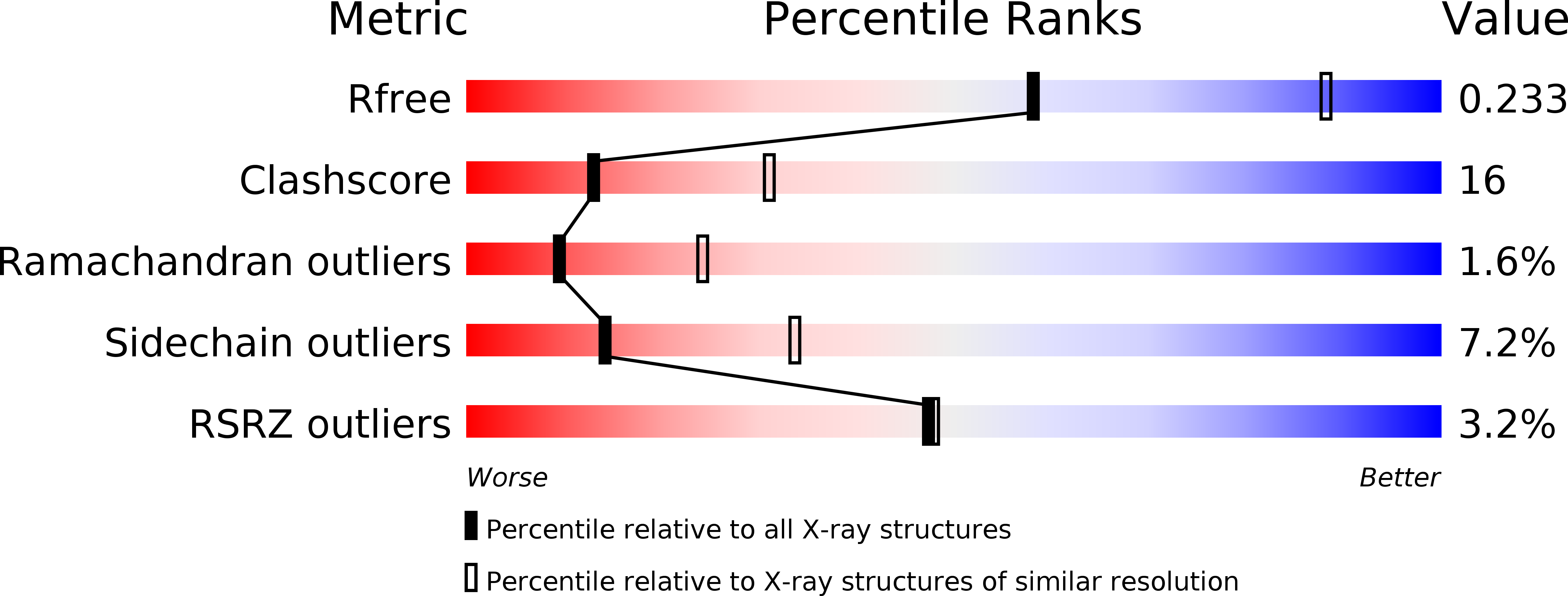
wwPDB Validation   3D Report Full Report


Ligand Structure Quality Assessment 


This is version 1.3 of the entry. See complete history


Literature

Structure and mechanism of mRNA cap (Guanine-n7) methyltransferase

Fabrega, C.Hausmann, S.Shen, V.Shuman, S.Lima, C.D.

(2004) Mol Cell 13: 77-89

  • DOI: https://doi.org/10.1016/s1097-2765(03)00522-7
  • Primary Citation of Related Structures:  
    1RI1, 1RI2, 1RI3, 1RI4, 1RI5

  • PubMed Abstract: 

    A suite of crystal structures is reported for a cellular mRNA cap (guanine-N7) methyltransferase in complex with AdoMet, AdoHcy, and the cap guanylate. Superposition of ligand complexes suggests an in-line mechanism of methyl transfer, albeit without direct contacts between the enzyme and either the N7 atom of guanine (the attacking nucleophile), the methyl carbon of AdoMet, or the sulfur of AdoMet/AdoHcy (the leaving group). The structures indicate that catalysis of cap N7 methylation is accomplished by optimizing proximity and orientation of the substrates, assisted by a favorable electrostatic environment. The enzyme-ligand structures, together with new mutational data, fully account for the biochemical specificity of the cap guanine-N7 methylation reaction, an essential and defining step of eukaryotic mRNA synthesis.


  • Organizational Affiliation

    Biochemistry Department, Structural Biology Program, Weill Medical College, Cornell University, New York, NY 10021, USA.


Macromolecules
Find similar proteins by:  (by identity cutoff)  |  3D Structure
Entity ID: 1
MoleculeChains Sequence LengthOrganismDetailsImage
mRNA CAPPING ENZYME298Encephalitozoon cuniculiMutation(s): 0 
Gene Names: ECU10_0380
EC: 2.1.1.56
UniProt
Find proteins for Q8SR66 (Encephalitozoon cuniculi (strain GB-M1))
Explore Q8SR66 
Go to UniProtKB:  Q8SR66
Entity Groups  
Sequence Clusters30% Identity50% Identity70% Identity90% Identity95% Identity100% Identity
UniProt GroupQ8SR66
Sequence Annotations
Expand
  • Reference Sequence
Small Molecules
Ligands 1 Unique
IDChains Name / Formula / InChI Key2D Diagram3D Interactions
GTG
Query on GTG

Download Ideal Coordinates CCD File 
B [auth A]7-METHYL-GUANOSINE-5'-TRIPHOSPHATE-5'-GUANOSINE
C21 H30 N10 O18 P3
FHHZHGZBHYYWTG-INFSMZHSSA-O
Experimental Data & Validation

Experimental Data

  • Method: X-RAY DIFFRACTION
  • Resolution: 2.70 Å
  • R-Value Free: 0.295 
  • R-Value Work: 0.224 
  • R-Value Observed: 0.224 
  • Space Group: P 31 2 1
Unit Cell:
Length ( Å )Angle ( ˚ )
a = 62.819α = 90
b = 62.819β = 90
c = 111.818γ = 120
Software Package:
Software NamePurpose
CNSrefinement
DENZOdata reduction
SCALEPACKdata scaling
SHARPphasing

Structure Validation

View Full Validation Report


Ligand Structure Quality Assessment 


Entry History 

Deposition Data

Revision History  (Full details and data files)

  • Version 1.0: 2004-02-03
    Type: Initial release
  • Version 1.1: 2008-04-29
    Changes: Version format compliance
  • Version 1.2: 2011-07-13
    Changes: Version format compliance
  • Version 1.3: 2024-02-14
    Changes: Data collection, Database references, Derived calculations