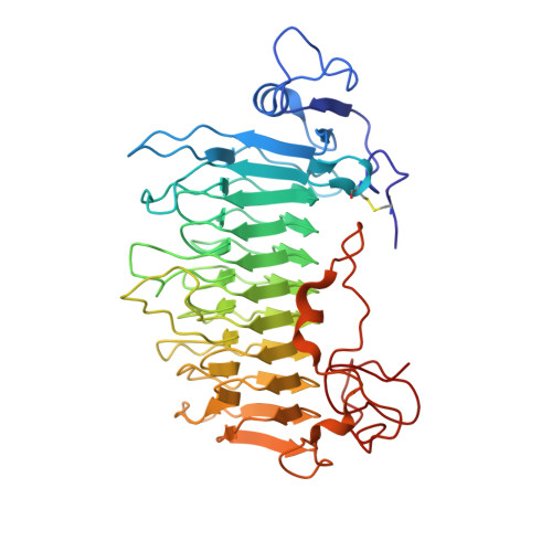
1RU4

Crystal structure of pectate lyase Pel9A


Experimental Data Snapshot

  • Method: X-RAY DIFFRACTION
  • Resolution: 1.60 Å
  • R-Value Free: 0.186 
  • R-Value Work: 0.167 
  • R-Value Observed: 0.177 

wwPDB Validation   3D Report Full Report


This is version 1.3 of the entry. See complete history


Literature

The crystal structure of pectate lyase Pel9A from Erwinia chrysanthemi

Jenkins, J.Shevchik, V.E.Hugouvieux-Cotte-Pattat, N.Pickersgill, R.W.

(2004) J Biol Chem 279: 9139-9145

  • DOI: https://doi.org/10.1074/jbc.M311390200
  • Primary Citation of Related Structures:  
    1RU4

  • PubMed Abstract: 

    The "family 9 polysaccharide lyase" pectate lyase L (Pel9A) from Erwinia chrysanthemi comprises a 10-coil parallel beta-helix domain with distinct structural features including an asparagine ladder and aromatic stack at novel positions within the superhelical structure. Pel9A has a single high affinity calcium-binding site strikingly similar to the "primary" calcium-binding site described previously for the family Pel1A pectate lyases, and there is strong evidence for a common second calcium ion that binds between enzyme and substrate in the "Michaelis" complex. Although the primary calcium ion binds substrate in subsite -1, it is the second calcium ion, whose binding site is formed by the coming together of enzyme and substrate, that facilitates abstraction of the C5 proton from the sacharride in subsite +1. The role of the second calcium is to withdraw electrons from the C6 carboxylate of the substrate, thereby acidifying the C5 proton facilitating its abstraction and resulting in an E1cb-like anti-beta-elimination mechanism. The active site geometries and mechanism of Pel1A and Pel9A are closely similar, but the catalytic base is a lysine in the Pel9A enzymes as opposed to an arginine in the Pel1A enzymes.


  • Organizational Affiliation

    Institute of Food Research, Norwich Research Park, Colney Lane, Norwich, NR4 7UA, United Kingdom.


Macromolecules
Find similar proteins by:  (by identity cutoff)  |  3D Structure
Entity ID: 1
MoleculeChains Sequence LengthOrganismDetailsImage
Pectate lyase400Dickeya chrysanthemiMutation(s): 0 
Gene Names: PelL
EC: 4.2.2.2
UniProt
Find proteins for P0C1A7 (Dickeya dadantii (strain 3937))
Explore P0C1A7 
Go to UniProtKB:  P0C1A7
Entity Groups  
Sequence Clusters30% Identity50% Identity70% Identity90% Identity95% Identity100% Identity
UniProt GroupP0C1A7
Sequence Annotations
Expand
  • Reference Sequence
Experimental Data & Validation

Experimental Data

  • Method: X-RAY DIFFRACTION
  • Resolution: 1.60 Å
  • R-Value Free: 0.186 
  • R-Value Work: 0.167 
  • R-Value Observed: 0.177 
  • Space Group: P 21 21 21
Unit Cell:
Length ( Å )Angle ( ˚ )
a = 55.33α = 90
b = 57.83β = 90
c = 114.93γ = 90
Software Package:
Software NamePurpose
DENZOdata reduction
SCALEPACKdata scaling
CCP4model building
CNSrefinement
REFMACrefinement
ARP/wARPmodel building
CCP4phasing

Structure Validation

View Full Validation Report


Entry History 

Deposition Data

Revision History  (Full details and data files)

  • Version 1.0: 2004-04-13
    Type: Initial release
  • Version 1.1: 2008-04-29
    Changes: Version format compliance
  • Version 1.2: 2011-07-13
    Changes: Version format compliance
  • Version 1.3: 2024-10-30
    Changes: Data collection, Database references, Derived calculations, Structure summary