1RYR

REPLICATION OF A CIS-SYN THYMINE DIMER AT ATOMIC RESOLUTION


Experimental Data Snapshot

  • Method: X-RAY DIFFRACTION
  • Resolution: 2.28 Å
  • R-Value Free: 0.281 
  • R-Value Work: 0.253 
  • R-Value Observed: 0.253 

wwPDB Validation   3D Report Full Report


Ligand Structure Quality Assessment 


This is version 1.4 of the entry. See complete history


Literature

Replication of a Cis-Syn Thymine Dimer at Atomic Resolution

Ling, H.Boudsocq, F.Plosky, B.Woodgate, R.Yang, W.

(2003) Nature 424: 1083-1087

  • DOI: https://doi.org/10.1038/nature01919
  • Primary Citation of Related Structures:  
    1RYR, 1RYS

  • PubMed Abstract: 

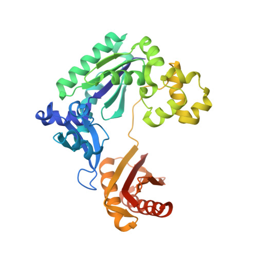
    Ultraviolet light damages DNA by catalysing covalent bond formation between adjacent pyrimidines, generating cis-syn cyclobutane pyrimidine dimers (CPDs) as the most common lesion. CPDs block DNA replication by high-fidelity DNA polymerases, but they can be efficiently bypassed by the Y-family DNA polymerase pol eta. Mutations in POLH encoding pol eta are implicated in nearly 20% of xeroderma pigmentosum, a human disease characterized by extreme sensitivity to sunlight and predisposition to skin cancer. Here we have determined two crystal structures of Dpo4, an archaeal pol eta homologue, complexed with CPD-containing DNA, where the 3' and 5' thymine of the CPD separately serves as a templating base. The 3' thymine of the CPD forms a Watson-Crick base pair with the incoming dideoxyATP, but the 5' thymine forms a Hoogsteen base pair with the dideoxyATP in syn conformation. Dpo4 retains a similar tertiary structure, but each unusual DNA structure is individually fitted into the active site for catalysis. A model of the pol eta-CPD complex built from the crystal structures of Saccharomyces cerevisiae apo-pol eta and the Dpo4-CPD complex suggests unique features that allow pol eta to efficiently bypass CPDs.


  • Organizational Affiliation

    Laboratory of Molecular Biology, National Institute of Diabetes and Digestive and Kidney Diseases, National Institutes of Health, Bethesda, Maryland 20892, USA.


Macromolecules

Find similar proteins by:  (by identity cutoff)  |  3D Structure
Entity ID: 3
MoleculeChains Sequence LengthOrganismDetailsImage
DNA polymerase IVC [auth A]352Saccharolobus solfataricusMutation(s): 0 
Gene Names: DPO4
EC: 2.7.7.7
UniProt
Find proteins for Q97W02 (Saccharolobus solfataricus (strain ATCC 35092 / DSM 1617 / JCM 11322 / P2))
Explore Q97W02 
Go to UniProtKB:  Q97W02
Entity Groups  
Sequence Clusters30% Identity50% Identity70% Identity90% Identity95% Identity100% Identity
UniProt GroupQ97W02
Sequence Annotations
Expand
  • Reference Sequence

Find similar nucleic acids by:  Sequence   |   3D Structure  

Entity ID: 1
MoleculeChains LengthOrganismImage
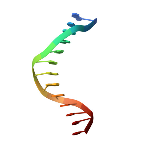
5'-D(*GP*GP*GP*GP*GP*AP*AP*GP*GP*AP*TP*TP*C)-3'A [auth B]13N/A
Sequence Annotations
Expand
  • Reference Sequence

Find similar nucleic acids by:  Sequence   |   3D Structure  

Entity ID: 2
MoleculeChains LengthOrganismImage
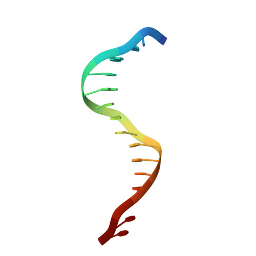
5'-D(*TP*TP*TP*GP*AP*AP*TP*CP*CP*TP*TP*CP*CP*CP*CP*C)-3'B [auth C]16N/A
Sequence Annotations
Expand
  • Reference Sequence
Experimental Data & Validation

Experimental Data

  • Method: X-RAY DIFFRACTION
  • Resolution: 2.28 Å
  • R-Value Free: 0.281 
  • R-Value Work: 0.253 
  • R-Value Observed: 0.253 
  • Space Group: P 21 21 2
Unit Cell:
Length ( Å )Angle ( ˚ )
a = 98.519α = 90
b = 101.556β = 90
c = 52.246γ = 90
Software Package:
Software NamePurpose
HKL-2000data collection
SCALEPACKdata scaling
CNSrefinement
HKL-2000data reduction
CNSphasing

Structure Validation

View Full Validation Report


Ligand Structure Quality Assessment 


Entry History 

Deposition Data

Revision History  (Full details and data files)

  • Version 1.0: 2004-02-10
    Type: Initial release
  • Version 1.1: 2008-04-29
    Changes: Version format compliance
  • Version 1.2: 2011-07-13
    Changes: Version format compliance
  • Version 1.3: 2018-04-04
    Changes: Data collection
  • Version 1.4: 2024-02-14
    Changes: Data collection, Database references, Derived calculations