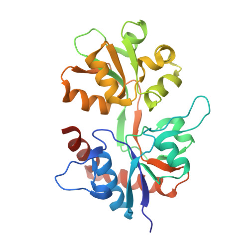
1TT1

CRYSTAL STRUCTURE OF THE GLUR6 LIGAND BINDING CORE IN COMPLEX WITH KAINATE 1.93 A RESOLUTION


Experimental Data Snapshot

  • Method: X-RAY DIFFRACTION
  • Resolution: 1.93 Å
  • R-Value Free: 0.216 
  • R-Value Work: 0.189 
  • R-Value Observed: 0.189 

Starting Model: experimental
View more details

wwPDB Validation   3D Report Full Report


Ligand Structure Quality Assessment 


This is version 1.4 of the entry. See complete history


Literature

Crystal structures of the GluR5 and GluR6 ligand binding cores: Molecular mechanisms underlying kainate receptor selectivity

Mayer, M.L.

(2005) Neuron 45: 539-552

  • DOI: https://doi.org/10.1016/j.neuron.2005.01.031
  • Primary Citation of Related Structures:  
    1S50, 1S7Y, 1S9T, 1SD3, 1TT1, 1TXF

  • PubMed Abstract: 

    Little is known about the molecular mechanisms underlying differences in the ligand binding properties of AMPA, kainate, and NMDA subtype glutamate receptors. Crystal structures of the GluR5 and GluR6 kainate receptor ligand binding cores in complexes with glutamate, 2S,4R-4-methylglutamate, kainate, and quisqualate have now been solved. The structures reveal that the ligand binding cavities are 40% (GluR5) and 16% (GluR6) larger than for GluR2. The binding of AMPA- and GluR5-selective agonists to GluR6 is prevented by steric occlusion, which also interferes with the high-affinity binding of 2S,4R-4-methylglutamate to AMPA receptors. Strikingly, the extent of domain closure produced by the GluR6 partial agonist kainate is only 3 degrees less than for glutamate and 11 degrees greater than for the GluR2 kainate complex. This, together with extensive interdomain contacts between domains 1 and 2 of GluR5 and GluR6, absent from AMPA receptors, likely contributes to the high stability of GluR5 and GluR6 kainate complexes.


  • Organizational Affiliation

    Porter Neuroscience Research Center, National Institute of Child Health and Human Development, National Institutes of Health, Department of Health and Human Services, Bethesda, Maryland 20892, USA. [email protected]


Macromolecules
Find similar proteins by:  (by identity cutoff)  |  3D Structure
Entity ID: 1
MoleculeChains Sequence LengthOrganismDetailsImage
Glutamate receptor, ionotropic kainate 2
A, B
259Rattus norvegicusMutation(s): 0 
Gene Names: GRIK2GLUR6
UniProt
Find proteins for P42260 (Rattus norvegicus)
Explore P42260 
Go to UniProtKB:  P42260
Entity Groups  
Sequence Clusters30% Identity50% Identity70% Identity90% Identity95% Identity100% Identity
UniProt GroupP42260
Sequence Annotations
Expand
  • Reference Sequence
Small Molecules
Binding Affinity Annotations 
IDSourceBinding Affinity
KAI PDBBind:  1TT1 IC50: 7 (nM) from 1 assay(s)
Experimental Data & Validation

Experimental Data

  • Method: X-RAY DIFFRACTION
  • Resolution: 1.93 Å
  • R-Value Free: 0.216 
  • R-Value Work: 0.189 
  • R-Value Observed: 0.189 
  • Space Group: P 1 21 1
Unit Cell:
Length ( Å )Angle ( ˚ )
a = 46.682α = 90
b = 106.228β = 101.38
c = 57.665γ = 90
Software Package:
Software NamePurpose
CNSrefinement
HKL-2000data reduction
SCALEPACKdata scaling
AMoREphasing

Structure Validation

View Full Validation Report


Ligand Structure Quality Assessment 


Entry History 

Deposition Data

  • Released Date: 2005-02-08 
  • Deposition Author(s): Mayer, M.L.

Revision History  (Full details and data files)

  • Version 1.0: 2005-02-08
    Type: Initial release
  • Version 1.1: 2008-04-01
    Changes: Version format compliance
  • Version 1.2: 2011-07-13
    Changes: Version format compliance
  • Version 1.3: 2017-07-26
    Changes: Advisory, Source and taxonomy
  • Version 1.4: 2023-08-23
    Changes: Advisory, Data collection, Database references, Derived calculations, Refinement description