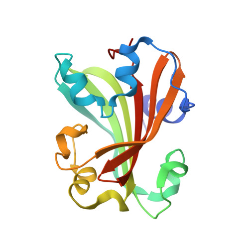
1TT8

CHORISMATE LYASE WITH PRODUCT, 1.0 A RESOLUTION


Experimental Data Snapshot

  • Method: X-RAY DIFFRACTION
  • Resolution: 1.00 Å
  • R-Value Free: 0.153 
  • R-Value Work: 0.123 
  • R-Value Observed: 0.123 

Starting Model: experimental
View more details

wwPDB Validation   3D Report Full Report


This is version 1.5 of the entry. See complete history


Literature

Structural analysis of ligand binding and catalysis in chorismate lyase.

Smith, N.Roitberg, A.E.Rivera, E.Howard, A.Holden, M.J.Mayhew, M.Kaistha, S.Gallagher, D.T.

(2006) Arch Biochem Biophys 445: 72-80

  • DOI: https://doi.org/10.1016/j.abb.2005.10.026
  • Primary Citation of Related Structures:  
    1JD3, 1TT8, 1XLR, 2AHC

  • PubMed Abstract: 

    Chorismate lyase (CL) removes the pyruvyl group from chorismate to provide 4-hydroxybenzoate (4HB) for the ubiquinone pathway. We previously reported the crystal structure at 1.4A resolution of the Escherichia coli CL with bound 4HB product, showing that the product is bound in an internal cavity behind two flaps. To provide a more complete basis for understanding CL's unusual ligand-binding properties and mechanism of action, we now report four crystal structures of CL mutants and inhibitor complexes, together with binding and activity measurements and molecular dynamics simulations. First, an ultrahigh resolution (1.0A) crystal structure of the CL*product complex reveals details of a substrate-sized internal cavity, also behind the flaps, near the product site. Second, a 2.4A structure of CL complexed with the inhibitor vanillate shows the flaps partly opened relative to their product-bound positions. Third, a 2.0A structure of the G90A mutant with bound product reveals the basis for tighter product binding and kinetic effects of this active site mutation. Fourth, the combination of the G90A mutation with the vanillate inhibitor produces a 1.9A structure containing two inhibitor molecules, one in the product site and the other in the adjacent cavity. The two sites are connected by a short tunnel that is partly open at each end, suggesting that CL may operate via a 2-site or tunnel mechanism.


  • Organizational Affiliation

    University of Maryland Biotechnology Institute, Rockville, MD 20850, USA.


Macromolecules
Find similar proteins by:  (by identity cutoff)  |  3D Structure
Entity ID: 1
MoleculeChains Sequence LengthOrganismDetailsImage
Chorismate-pyruvate lyase164Escherichia coliMutation(s): 2 
Gene Names: ubiCb4039
EC: 4 (PDB Primary Data), 4.1.3.40 (UniProt)
UniProt
Find proteins for P26602 (Escherichia coli (strain K12))
Explore P26602 
Go to UniProtKB:  P26602
Entity Groups  
Sequence Clusters30% Identity50% Identity70% Identity90% Identity95% Identity100% Identity
UniProt GroupP26602
Sequence Annotations
Expand
  • Reference Sequence
Small Molecules
Ligands 1 Unique
IDChains Name / Formula / InChI Key2D Diagram3D Interactions
PHB
Query on PHB

Download Ideal Coordinates CCD File 
B [auth A]P-HYDROXYBENZOIC ACID
C7 H6 O3
FJKROLUGYXJWQN-UHFFFAOYSA-N
Experimental Data & Validation

Experimental Data

  • Method: X-RAY DIFFRACTION
  • Resolution: 1.00 Å
  • R-Value Free: 0.153 
  • R-Value Work: 0.123 
  • R-Value Observed: 0.123 
  • Space Group: P 1
Unit Cell:
Length ( Å )Angle ( ˚ )
a = 31.315α = 67.57
b = 33.849β = 87.83
c = 39.776γ = 65.66
Software Package:
Software NamePurpose
X-GENdata scaling
X-GENdata reduction
SHELXmodel building
SHELXL-97refinement
SHELXphasing

Structure Validation

View Full Validation Report


Entry History 

Deposition Data

Revision History  (Full details and data files)

  • Version 1.0: 2004-12-28
    Type: Initial release
  • Version 1.1: 2008-04-30
    Changes: Version format compliance
  • Version 1.2: 2011-07-13
    Changes: Version format compliance
  • Version 1.3: 2017-10-11
    Changes: Refinement description
  • Version 1.4: 2021-10-27
    Changes: Database references, Derived calculations
  • Version 1.5: 2023-08-23
    Changes: Data collection, Refinement description