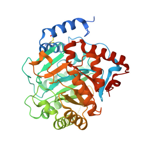
1TV5

Plasmodium falciparum dihydroorotate dehydrogenase with a bound inhibitor


Experimental Data Snapshot

  • Method: X-RAY DIFFRACTION
  • Resolution: 2.40 Å
  • R-Value Free: 0.243 
  • R-Value Work: 0.201 
  • R-Value Observed: 0.201 

Starting Model: experimental
View more details

wwPDB Validation   3D Report Full Report


Ligand Structure Quality Assessment 


This is version 1.5 of the entry. See complete history


Literature

Structure of Plasmodium falciparum dihydroorotate dehydrogenase with a bound inhibitor.

Hurt, D.E.Widom, J.Clardy, J.

(2006) Acta Crystallogr D Biol Crystallogr 62: 312-323

  • DOI: https://doi.org/10.1107/S0907444905042642
  • Primary Citation of Related Structures:  
    1TV5

  • PubMed Abstract: 

    Membrane-associated dihydroorotate dehydrogenase (DHODH) is an antimalarial therapeutic target without an effective inhibitor. Studies on human DHODH (HsDHODH) led to a structural mechanistic model in which respiratory quinones bind in a tunnel formed by the highly variable N-terminus that leads to the flavin mononucleotide-binding site. The therapeutic agents leflunomide (Arava) and brequinar sodium inhibit HsDHODH by binding in this tunnel. Plasmodium falciparum DHODH (PfDHODH) and HsDHODH have markedly different sensitivities to the two drugs. To understand the structural basis of this differential sensitivity and begin a structure-based drug-design cycle for PfDHODH inhibitors, the three-dimensional structure (2.4 Angstroms, R = 20.1%) of PfDHODH bound to the active metabolite of leflunomide was determined by X-ray crystallography. Comparison of the structures of HsDHODH and PfDHODH reveals a completely different binding mode for the same inhibitor in these two catalytically identical enzymes and explains the previously observed species-specific preferential binding. Because no effective inhibitors have been described for PfDHODH, this structure provides critical insight for the design of potential antimalarials.


  • Organizational Affiliation

    Department of Chemistry and Chemical Biology, Cornell University, Ithaca, NY 14850, USA.


Macromolecules
Find similar proteins by:  (by identity cutoff)  |  3D Structure
Entity ID: 1
MoleculeChains Sequence LengthOrganismDetailsImage
Dihydroorotate dehydrogenase homolog, mitochondrial443Plasmodium falciparumMutation(s): 0 
Gene Names: dhod
EC: 1.3.99.11 (PDB Primary Data), 1.3.3.1 (PDB Primary Data), 1.3.5.2 (UniProt)
UniProt
Find proteins for Q08210 (Plasmodium falciparum (isolate 3D7))
Explore Q08210 
Go to UniProtKB:  Q08210
Entity Groups  
Sequence Clusters30% Identity50% Identity70% Identity90% Identity95% Identity100% Identity
UniProt GroupQ08210
Sequence Annotations
Expand
  • Reference Sequence
Small Molecules
Ligands 5 Unique
IDChains Name / Formula / InChI Key2D Diagram3D Interactions
FMN
Query on FMN

Download Ideal Coordinates CCD File 
D [auth A]FLAVIN MONONUCLEOTIDE
C17 H21 N4 O9 P
FVTCRASFADXXNN-SCRDCRAPSA-N
N8E
Query on N8E

Download Ideal Coordinates CCD File 
F [auth A],
G [auth A]
3,6,9,12,15-PENTAOXATRICOSAN-1-OL
C18 H38 O6
MJELOWOAIAAUJT-UHFFFAOYSA-N
A26
Query on A26

Download Ideal Coordinates CCD File 
C [auth A](2Z)-2-cyano-3-hydroxy-N-[4-(trifluoromethyl)phenyl]but-2-enamide
C12 H9 F3 N2 O2
UTNUDOFZCWSZMS-YFHOEESVSA-N
ORO
Query on ORO

Download Ideal Coordinates CCD File 
E [auth A]OROTIC ACID
C5 H4 N2 O4
PXQPEWDEAKTCGB-UHFFFAOYSA-N
SO4
Query on SO4

Download Ideal Coordinates CCD File 
B [auth A]SULFATE ION
O4 S
QAOWNCQODCNURD-UHFFFAOYSA-L
Binding Affinity Annotations 
IDSourceBinding Affinity
A26 BindingDB:  1TV5 IC50: 1.91e+5 (nM) from 1 assay(s)
Experimental Data & Validation

Experimental Data

  • Method: X-RAY DIFFRACTION
  • Resolution: 2.40 Å
  • R-Value Free: 0.243 
  • R-Value Work: 0.201 
  • R-Value Observed: 0.201 
  • Space Group: H 3 2
Unit Cell:
Length ( Å )Angle ( ˚ )
a = 105.428α = 90
b = 105.428β = 90
c = 276.632γ = 120
Software Package:
Software NamePurpose
DENZOdata reduction
SCALAdata scaling
RESOLVEmodel building
CNSrefinement
CCP4data scaling
RESOLVEphasing

Structure Validation

View Full Validation Report


Ligand Structure Quality Assessment 


Entry History 

Deposition Data

Revision History  (Full details and data files)

  • Version 1.0: 2005-09-06
    Type: Initial release
  • Version 1.1: 2008-04-30
    Changes: Version format compliance
  • Version 1.2: 2011-07-13
    Changes: Derived calculations, Version format compliance
  • Version 1.3: 2012-07-18
    Changes: Non-polymer description
  • Version 1.4: 2023-08-23
    Changes: Data collection, Database references, Derived calculations, Refinement description
  • Version 1.5: 2024-10-09
    Changes: Structure summary