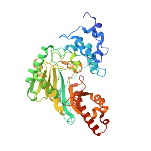
1VRP

The 2.1 Structure of T. californica Creatine Kinase Complexed with the Transition-State Analogue Complex, ADP-Mg 2+ /NO3-/Creatine


Experimental Data Snapshot

  • Method: X-RAY DIFFRACTION
  • Resolution: 2.10 Å
  • R-Value Free: 0.278 
  • R-Value Work: 0.242 
  • R-Value Observed: 0.260 

Starting Model: experimental
View more details

wwPDB Validation   3D Report Full Report


Ligand Structure Quality Assessment 


This is version 1.3 of the entry. See complete history


Literature

The 2.1 A Structure of Torpedo californica Creatine Kinase Complexed with the ADP-Mg(2+)-NO3(-)-Creatine Transition-State Analogue Complex

Lahiri, S.D.Wang, P.F.Babbitt, P.C.McLeish, M.J.Kenyon, G.L.Allen, K.N.

(2002) Biochemistry 41: 13861-13867

  • DOI: https://doi.org/10.1021/bi026655p
  • Primary Citation of Related Structures:  
    1VRP

  • PubMed Abstract: 

    Creatine kinase (CK) catalyzes the reversible conversion of creatine and ATP to phosphocreatine and ADP, thereby helping maintain energy homeostasis in the cell. Here we report the first X-ray structure of CK bound to a transition-state analogue complex (CK-TSAC). Cocrystallization of the enzyme from Torpedo californica (TcCK) with ADP-Mg(2+), nitrate, and creatine yielded a homodimer, one monomer of which was liganded to a TSAC complex while the second monomer was bound to ADP-Mg(2+) alone. The structures of both monomers were determined to 2.1 A resolution. The creatine is located with the guanidino nitrogen cis to the methyl group positioned to perform in-line attack at the gamma-phosphate of ATP-Mg(2+), while the ADP-Mg(2+) is in a conformation similar to that found in the TSAC-bound structure of the homologue arginine kinase (AK). Three ligands to Mg(2+) are contributed by ADP and nitrate and three by ordered water molecules. The most striking difference between the substrate-bound and TSAC-bound structures is the movement of two loops, comprising residues 60-70 and residues 323-332. In the TSAC-bound structure, both loops move into the active site, resulting in the positioning of two hydrophobic residues (one from each loop), Ile69 and Val325, near the methyl group of creatine. This apparently provides a specificity pocket for optimal creatine binding as this interaction is missing in the AK structure. In addition, the active site of the transition-state analogue complex is completely occluded from solvent, unlike the ADP-Mg(2+)-bound monomer and the unliganded structures reported previously.


  • Organizational Affiliation

    Department of Physiology and Biophysics, Boston University School of Medicine, Boston, Massachusetts 02155, USA.


Macromolecules
Find similar proteins by:  (by identity cutoff)  |  3D Structure
Entity ID: 1
MoleculeChains Sequence LengthOrganismDetailsImage
Creatine Kinase, M chain
A, B
381Tetronarce californicaMutation(s): 0 
Gene Names: FSCCKPA
EC: 2.7.3.2
UniProt
Find proteins for P04414 (Tetronarce californica)
Explore P04414 
Go to UniProtKB:  P04414
Entity Groups  
Sequence Clusters30% Identity50% Identity70% Identity90% Identity95% Identity100% Identity
UniProt GroupP04414
Sequence Annotations
Expand
  • Reference Sequence
Small Molecules
Ligands 4 Unique
IDChains Name / Formula / InChI Key2D Diagram3D Interactions
ADP
Query on ADP

Download Ideal Coordinates CCD File 
D [auth A],
G [auth B]
ADENOSINE-5'-DIPHOSPHATE
C10 H15 N5 O10 P2
XTWYTFMLZFPYCI-KQYNXXCUSA-N
IOM
Query on IOM

Download Ideal Coordinates CCD File 
H [auth B](DIAMINOMETHYL-METHYL-AMINO)-ACETIC ACID
C4 H11 N3 O2
YNHURFGTTODJOO-UHFFFAOYSA-N
NO3
Query on NO3

Download Ideal Coordinates CCD File 
F [auth B]NITRATE ION
N O3
NHNBFGGVMKEFGY-UHFFFAOYSA-N
MG
Query on MG

Download Ideal Coordinates CCD File 
C [auth A],
E [auth B]
MAGNESIUM ION
Mg
JLVVSXFLKOJNIY-UHFFFAOYSA-N
Experimental Data & Validation

Experimental Data

  • Method: X-RAY DIFFRACTION
  • Resolution: 2.10 Å
  • R-Value Free: 0.278 
  • R-Value Work: 0.242 
  • R-Value Observed: 0.260 
  • Space Group: P 21 21 21
Unit Cell:
Length ( Å )Angle ( ˚ )
a = 70.634α = 90
b = 87.189β = 90
c = 127.596γ = 90
Software Package:
Software NamePurpose
DENZOdata reduction
SCALEPACKdata scaling
AMoREphasing
CNSrefinement

Structure Validation

View Full Validation Report


Ligand Structure Quality Assessment 


Entry History 

Deposition Data

Revision History  (Full details and data files)

  • Version 1.0: 2005-05-03
    Type: Initial release
  • Version 1.1: 2008-04-27
    Changes: Version format compliance
  • Version 1.2: 2011-07-13
    Changes: Version format compliance
  • Version 1.3: 2023-08-23
    Changes: Data collection, Database references, Derived calculations, Refinement description