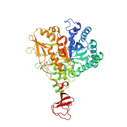
1W1Y

Crystal structure of S. marcescens chitinase B in complex with the cyclic dipeptide inhibitor cyclo-(L-Tyr-L-Pro) at 1.85 A resolution


Experimental Data Snapshot

  • Method: X-RAY DIFFRACTION
  • Resolution: 1.85 Å
  • R-Value Free: 0.202 
  • R-Value Work: 0.170 
  • R-Value Observed: 0.170 

Starting Model: experimental
View more details

wwPDB Validation   3D Report Full Report


Ligand Structure Quality Assessment 


This is version 1.4 of the entry. See complete history


Literature

Structure-Based Exploration of Cyclic Dipeptide Chitinase Inhibitors

Houston, D.R.Synstad, B.Eijsink, V.G.H.Stark, M.J.Eggleston, I.Van Aalten, D.M.F.

(2004) J Med Chem 47: 5713

  • DOI: https://doi.org/10.1021/jm049940a
  • Primary Citation of Related Structures:  
    1W1P, 1W1T, 1W1V, 1W1Y

  • PubMed Abstract: 

    Family 18 chitinases play an essential role in a range of pathogens and pests. Several inhibitors are known, including the potent inhibitors argadin and allosamidin, and the structures of these in complex with chitinases have been elucidated. Recent structural analysis has revealed that CI-4 [cyclo-(L-Arg-D-Pro)] inhibits family 18 chitinases by mimicking the structure of the proposed reaction intermediate. Here we report the high-resolution structures of four new CI-4 derivatives, cyclo-(L-Arg-L-Pro), cyclo-(Gly-L-Pro), cyclo-(L-His-L-Pro), and cyclo-(L-Tyr-L-Pro), in complex with a family 18 chitinase. In addition, details of enzyme inhibition and in vivo activity against Saccharomyces cerevisiae are presented. The structures reveal that the common cyclo-(Gly-Pro) substructure is sufficient for binding, allowing modification of the side chain of the nonproline residue. This suggests that design of cyclic dipeptides with a view to increasing inhibition of family 18 chitinases should be possible through relatively accessible chemistry. The derivatives presented here in complex with chitinase B from Serratia marcescens provide further insight into the mechanism of inhibition of chitinases by cyclic dipeptides as well as providing a new scaffold for chitinase inhibitor design.


  • Organizational Affiliation

    Division of Biological Chemistry and Molecular Microbiology, School of Life Sciences, University of Dundee, Dundee DD1 5EH, Scotland, UK.


Macromolecules
Find similar proteins by:  (by identity cutoff)  |  3D Structure
Entity ID: 1
MoleculeChains Sequence LengthOrganismDetailsImage
CHITINASE B
A, B
499Serratia marcescensMutation(s): 0 
EC: 3.2.1.14
UniProt
Find proteins for Q54276 (Serratia marcescens)
Explore Q54276 
Go to UniProtKB:  Q54276
Entity Groups  
Sequence Clusters30% Identity50% Identity70% Identity90% Identity95% Identity100% Identity
UniProt GroupQ54276
Sequence Annotations
Expand
  • Reference Sequence
Small Molecules
Ligands 3 Unique
IDChains Name / Formula / InChI Key2D Diagram3D Interactions
TYP
Query on TYP

Download Ideal Coordinates CCD File 
K [auth A]
L [auth A]
M [auth A]
T [auth B]
U [auth B]
K [auth A],
L [auth A],
M [auth A],
T [auth B],
U [auth B],
V [auth B],
W [auth B]
CYCLO-(L-TYROSINE-L-PROLINE) INHIBITOR
C14 H16 N2 O3
LSGOTAXPWMCUCK-RYUDHWBXSA-N
SO4
Query on SO4

Download Ideal Coordinates CCD File 
I [auth A],
J [auth A]
SULFATE ION
O4 S
QAOWNCQODCNURD-UHFFFAOYSA-L
GOL
Query on GOL

Download Ideal Coordinates CCD File 
C [auth A]
D [auth A]
E [auth A]
F [auth A]
G [auth A]
C [auth A],
D [auth A],
E [auth A],
F [auth A],
G [auth A],
H [auth A],
N [auth B],
O [auth B],
P [auth B],
Q [auth B],
R [auth B],
S [auth B]
GLYCEROL
C3 H8 O3
PEDCQBHIVMGVHV-UHFFFAOYSA-N
Binding Affinity Annotations 
IDSourceBinding Affinity
TYP PDBBind:  1W1Y IC50: 2.40e+6 (nM) from 1 assay(s)
Experimental Data & Validation

Experimental Data

  • Method: X-RAY DIFFRACTION
  • Resolution: 1.85 Å
  • R-Value Free: 0.202 
  • R-Value Work: 0.170 
  • R-Value Observed: 0.170 
  • Space Group: P 21 21 21
Unit Cell:
Length ( Å )Angle ( ˚ )
a = 55.433α = 90
b = 103.528β = 90
c = 183.859γ = 90
Software Package:
Software NamePurpose
CNSrefinement
DENZOdata reduction
SCALEPACKdata scaling
CNSphasing

Structure Validation

View Full Validation Report


Ligand Structure Quality Assessment 


Entry History 

Deposition Data

Revision History  (Full details and data files)

  • Version 1.0: 2005-01-10
    Type: Initial release
  • Version 1.1: 2011-05-08
    Changes: Version format compliance
  • Version 1.2: 2011-07-13
    Changes: Version format compliance
  • Version 1.3: 2023-12-13
    Changes: Data collection, Database references, Other, Refinement description
  • Version 1.4: 2024-11-06
    Changes: Structure summary