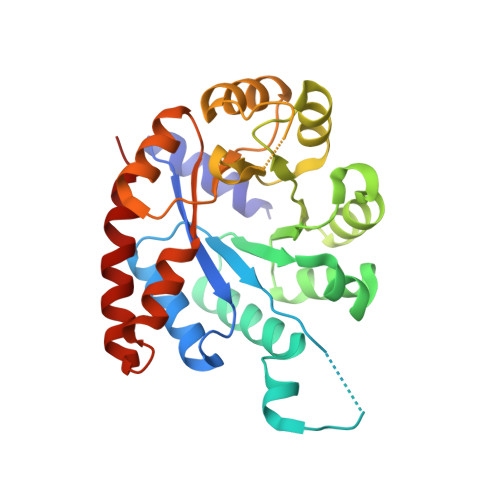
1WQ5

Crystal structure of tryptophan synthase alpha-subunit from Escherichia coli


Experimental Data Snapshot

  • Method: X-RAY DIFFRACTION
  • Resolution: 2.30 Å
  • R-Value Free: 0.248 
  • R-Value Work: 0.185 
  • R-Value Observed: 0.185 

Starting Model: experimental
View more details

wwPDB Validation   3D Report Full Report


This is version 1.4 of the entry. See complete history


Literature

Conformational Changes in the alpha-Subunit Coupled to Binding of the beta(2)-Subunit of Tryptophan Synthase from Escherichia coli: Crystal Structure of the Tryptophan Synthase alpha-Subunit Alon

Nishio, K.Morimoto, Y.Ishizuka, M.Ogasahara, K.Tsukihara, T.Yutani, K.

(2005) Biochemistry 44: 1184-1192

  • DOI: https://doi.org/10.1021/bi047927m
  • Primary Citation of Related Structures:  
    1V7Y, 1WQ5

  • PubMed Abstract: 

    When the tryptophan synthase alpha- and beta(2)-subunits combine to form the alpha(2)beta(2)-complex, the enzymatic activity of each subunit is stimulated by 1-2 orders of magnitude. To elucidate the structural basis of this mutual activation, it is necessary to determine the structures of the alpha- and beta-subunits alone and together with the alpha(2)beta(2)-complex. The crystal structures of the tryptophan synthase alpha(2)beta(2)-complex from Salmonella typhimurium (Stalpha(2)beta(2)-complex) have already been reported. However, the structures of the subunit alone from mesophiles have not yet been determined. The structure of the tryptophan synthase alpha-subunit alone from Escherichia coli (Ecalpha-subunit) was determined by an X-ray crystallographic analysis at 2.3 A, which is the first report on the subunits alone from the mesophiles. The biggest difference between the structures of the Ecalpha-subunit alone and the alpha-subunit in the Stalpha(2)beta(2)-complex (Stalpha-subunit) was as follows. Helix 2' in the Stalpha-subunit, including an active site residue (Asp60), was changed to a flexible loop in the Ecalpha-subunit alone. The conversion of the helix to a loop resulted in the collapse of the correct active site conformation. This region is also an important part for the mutual activation in the Stalpha(2)beta(2)-complex and interaction with the beta-subunit. These results suggest that the formation of helix 2'that is essential for the stimulation of the enzymatic activity of the alpha-subunit is constructed by the induced-fit mode involved in conformational changes upon interaction between the alpha- and beta-subunits. This also confirms the prediction of the conformational changes based on the thermodynamic analysis for the association between the alpha- and beta-subunits.


  • Organizational Affiliation

    Institute for Protein Research, Osaka University, 3-2 Yamadaoka, Suita, Osaka 565-0871, Japan.


Macromolecules
Find similar proteins by:  (by identity cutoff)  |  3D Structure
Entity ID: 1
MoleculeChains Sequence LengthOrganismDetailsImage
Tryptophan synthase alpha chain
A, B
268Escherichia coliMutation(s): 0 
EC: 4.2.1.20
UniProt
Find proteins for P0A877 (Escherichia coli (strain K12))
Explore P0A877 
Go to UniProtKB:  P0A877
Entity Groups  
Sequence Clusters30% Identity50% Identity70% Identity90% Identity95% Identity100% Identity
UniProt GroupP0A877
Sequence Annotations
Expand
  • Reference Sequence
Small Molecules
Ligands 2 Unique
IDChains Name / Formula / InChI Key2D Diagram3D Interactions
SO4
Query on SO4

Download Ideal Coordinates CCD File 
C [auth A]
D [auth A]
E [auth A]
F [auth A]
G [auth A]
C [auth A],
D [auth A],
E [auth A],
F [auth A],
G [auth A],
H [auth A],
I [auth A],
P [auth B],
Q [auth B],
R [auth B],
S [auth B],
T [auth B],
U [auth B]
SULFATE ION
O4 S
QAOWNCQODCNURD-UHFFFAOYSA-L
GOL
Query on GOL

Download Ideal Coordinates CCD File 
J [auth A]
K [auth A]
L [auth A]
M [auth A]
N [auth A]
J [auth A],
K [auth A],
L [auth A],
M [auth A],
N [auth A],
O [auth A],
V [auth B],
W [auth B]
GLYCEROL
C3 H8 O3
PEDCQBHIVMGVHV-UHFFFAOYSA-N
Experimental Data & Validation

Experimental Data

  • Method: X-RAY DIFFRACTION
  • Resolution: 2.30 Å
  • R-Value Free: 0.248 
  • R-Value Work: 0.185 
  • R-Value Observed: 0.185 
  • Space Group: C 1 2 1
Unit Cell:
Length ( Å )Angle ( ˚ )
a = 156.774α = 90
b = 44.626β = 96.46
c = 71.716γ = 90
Software Package:
Software NamePurpose
CNSrefinement
DENZOdata reduction
SCALEPACKdata scaling
CNSphasing

Structure Validation

View Full Validation Report


Entry History 

Revision History  (Full details and data files)

  • Version 1.0: 2005-02-15
    Type: Initial release
  • Version 1.1: 2008-04-30
    Changes: Version format compliance
  • Version 1.2: 2011-07-13
    Changes: Derived calculations, Version format compliance
  • Version 1.3: 2024-03-13
    Changes: Data collection, Database references, Derived calculations
  • Version 1.4: 2024-04-03
    Changes: Refinement description