1WYG

Crystal Structure of a Rat Xanthine Dehydrogenase Triple Mutant (C535A, C992R and C1324S)


Experimental Data Snapshot

  • Method: X-RAY DIFFRACTION
  • Resolution: 2.60 Å
  • R-Value Free: 0.248 
  • R-Value Work: 0.203 

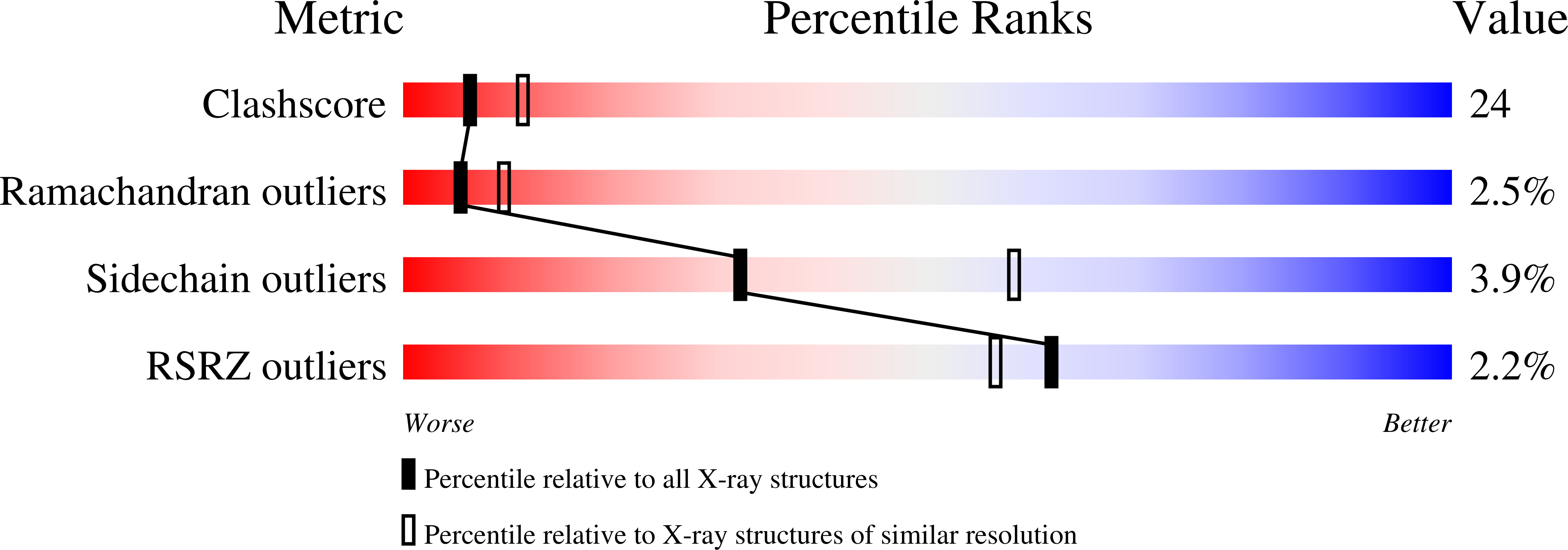
wwPDB Validation   3D Report Full Report


Ligand Structure Quality Assessment 


This is version 1.4 of the entry. See complete history


Literature

Mechanism of the Conversion of Xanthine Dehydrogenase to Xanthine Oxidase: IDENTIFICATION OF THE TWO CYSTEINE DISULFIDE BONDS AND CRYSTAL STRUCTURE OF A NON-CONVERTIBLE RAT LIVER XANTHINE DEHYDROGENASE MUTANT

Nishino, T.Okamoto, K.Kawaguchi, Y.Hori, H.Matsumura, T.Eger, B.T.Pai, E.F.Nishino, T.

(2005) J Biol Chem 280: 24888-24894

  • DOI: https://doi.org/10.1074/jbc.M501830200
  • Primary Citation of Related Structures:  
    1WYG

  • PubMed Abstract: 

    Mammalian xanthine dehydrogenase can be converted to xanthine oxidase by modification of cysteine residues or by proteolysis of the enzyme polypeptide chain. Here we present evidence that the Cys(535) and Cys(992) residues of rat liver enzyme are indeed involved in the rapid conversion from the dehydrogenase to the oxidase. The purified mutants C535A and/or C992R were significantly resistant to conversion by incubation with 4,4'-dithiodipyridine, whereas the recombinant wild-type enzyme converted readily to the oxidase type, indicating that these residues are responsible for the rapid conversion. The C535A/C992R mutant, however, converted very slowly during prolonged incubation with 4,4'-dithiodipyridine, and this slow conversion was blocked by the addition of NADH, suggesting that another cysteine couple located near the NAD(+) binding site is responsible for the slower conversion. On the other hand, the C535A/C992R/C1316S and C535A/C992R/C1324S mutants were completely resistant to conversion, even on prolonged incubation with 4,4'-dithiodipyridine, indicating that Cys(1316) and Cys(1324) are responsible for the slow conversion. The crystal structure of the C535A/C992R/C1324S mutant was determined in its demolybdo form, confirming its dehydrogenase conformation.


  • Organizational Affiliation

    Department of Biochemistry and Molecular Biology, Nippon Medical School, 1-1-5 Sendagi, Bunkyo-ku, Tokyo 113-8602, Japan. [email protected]


Macromolecules
Find similar proteins by:  (by identity cutoff)  |  3D Structure
Entity ID: 1
MoleculeChains Sequence LengthOrganismDetailsImage
Xanthine dehydrogenase/oxidase1,331Rattus norvegicusMutation(s): 3 
EC: 1.1.1.204 (PDB Primary Data), 1.1.3.22 (PDB Primary Data), 1.17.1.4 (UniProt), 1.17.3.2 (UniProt)
UniProt
Find proteins for P22985 (Rattus norvegicus)
Explore P22985 
Go to UniProtKB:  P22985
Entity Groups  
Sequence Clusters30% Identity50% Identity70% Identity90% Identity95% Identity100% Identity
UniProt GroupP22985
Sequence Annotations
Expand
  • Reference Sequence
Small Molecules
Ligands 6 Unique
IDChains Name / Formula / InChI Key2D Diagram3D Interactions
FAD
Query on FAD

Download Ideal Coordinates CCD File 
H [auth A]FLAVIN-ADENINE DINUCLEOTIDE
C27 H33 N9 O15 P2
VWWQXMAJTJZDQX-UYBVJOGSSA-N
FES
Query on FES

Download Ideal Coordinates CCD File 
F [auth A],
G [auth A]
FE2/S2 (INORGANIC) CLUSTER
Fe2 S2
NIXDOXVAJZFRNF-UHFFFAOYSA-N
SAL
Query on SAL

Download Ideal Coordinates CCD File 
I [auth A]2-HYDROXYBENZOIC ACID
C7 H6 O3
YGSDEFSMJLZEOE-UHFFFAOYSA-N
PO4
Query on PO4

Download Ideal Coordinates CCD File 
B [auth A],
C [auth A],
D [auth A]
PHOSPHATE ION
O4 P
NBIIXXVUZAFLBC-UHFFFAOYSA-K
ACY
Query on ACY

Download Ideal Coordinates CCD File 
J [auth A]ACETIC ACID
C2 H4 O2
QTBSBXVTEAMEQO-UHFFFAOYSA-N
CA
Query on CA

Download Ideal Coordinates CCD File 
E [auth A]CALCIUM ION
Ca
BHPQYMZQTOCNFJ-UHFFFAOYSA-N
Binding Affinity Annotations 
IDSourceBinding Affinity
SAL BindingDB:  1WYG Ki: 1.00e+8 (nM) from 1 assay(s)
Experimental Data & Validation

Experimental Data

  • Method: X-RAY DIFFRACTION
  • Resolution: 2.60 Å
  • R-Value Free: 0.248 
  • R-Value Work: 0.203 
  • Space Group: I 41 2 2
Unit Cell:
Length ( Å )Angle ( ˚ )
a = 134.253α = 90
b = 134.253β = 90
c = 523.315γ = 90
Software Package:
Software NamePurpose
HKL-2000data collection
SCALEPACKdata scaling
EPMRphasing
CNSrefinement
HKL-2000data reduction

Structure Validation

View Full Validation Report


Ligand Structure Quality Assessment 


Entry History 

Deposition Data

Revision History  (Full details and data files)

  • Version 1.0: 2005-05-31
    Type: Initial release
  • Version 1.1: 2008-04-30
    Changes: Version format compliance
  • Version 1.2: 2011-07-13
    Changes: Derived calculations, Version format compliance
  • Version 1.3: 2021-11-10
    Changes: Database references, Derived calculations
  • Version 1.4: 2024-05-29
    Changes: Data collection