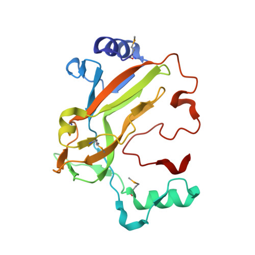
1XS4

dCTP deaminase from Escherichia coli- E138A mutant enzyme in complex with dCTP


Experimental Data Snapshot

  • Method: X-RAY DIFFRACTION
  • Resolution: 2.53 Å
  • R-Value Free: 0.225 
  • R-Value Work: 0.185 
  • R-Value Observed: 0.187 

Starting Model: experimental
View more details

wwPDB Validation   3D Report Full Report


Ligand Structure Quality Assessment 


This is version 1.6 of the entry. See complete history


Literature

Structures of dCTP deaminase from Escherichia coli with bound substrate and product: reaction mechanism and determinants of mono- and bifunctionality for a family of enzymes

Johansson, E.Fano, M.Bynck, J.H.Neuhard, J.Larsen, S.Sigurskjold, B.W.Christensen, U.Willemoes, M.

(2005) J Biol Chem 280: 3051-3059

  • DOI: https://doi.org/10.1074/jbc.M409534200
  • Primary Citation of Related Structures:  
    1XS1, 1XS4, 1XS6

  • PubMed Abstract: 

    dCTP deaminase (EC 3.5.4.13) catalyzes the deamination of dCTP forming dUTP that via dUTPase is the main pathway providing substrate for thymidylate synthase in Escherichia coli and Salmonella typhimurium. dCTP deaminase is unique among nucleoside and nucleotide deaminases as it functions without aid from a catalytic metal ion that facilitates preparation of a water molecule for nucleophilic attack on the substrate. Two active site amino acid residues, Arg(115) and Glu(138), were identified by mutational analysis as important for activity in E. coli dCTP deaminase. None of the mutant enzymes R115A, E138A, or E138Q had any detectable activity but circular dichroism spectra for all mutant enzymes were similar to wild type suggesting that the overall structure was not changed. The crystal structures of wild-type E. coli dCTP deaminase and the E138A mutant enzyme have been determined in complex with dUTP and Mg(2+), and the mutant enzyme also with the substrate dCTP and Mg(2+). The enzyme is a third member of the family of the structurally related trimeric dUTPases and the bifunctional dCTP deaminase-dUTPase from Methanocaldococcus jannaschii. However, the C-terminal fold is completely different from dUTPases resulting in an active site built from residues from two of the trimer subunits, and not from three subunits as in dUTPases. The nucleotides are well defined as well as Mg(2+) that is tridentately coordinated to the nucleotide phosphate chains. We suggest a catalytic mechanism for the dCTP deaminase and identify structural differences to dUTPases that prevent hydrolysis of the dCTP triphosphate.


  • Organizational Affiliation

    Centre for Crystallographic Studies, Department of Chemistry, University of Copenhagen Universitetsparken 5, DK-2100, Copenhagen, Denmark.


Macromolecules
Find similar proteins by:  (by identity cutoff)  |  3D Structure
Entity ID: 1
MoleculeChains Sequence LengthOrganismDetailsImage
Deoxycytidine triphosphate deaminase
A, B, C, D, E
A, B, C, D, E, F
193Escherichia coliMutation(s): 5 
Gene Names: dcd
EC: 3.5.4.13
UniProt
Find proteins for P28248 (Escherichia coli (strain K12))
Explore P28248 
Go to UniProtKB:  P28248
Entity Groups  
Sequence Clusters30% Identity50% Identity70% Identity90% Identity95% Identity100% Identity
UniProt GroupP28248
Sequence Annotations
Expand
  • Reference Sequence
Small Molecules
Ligands 2 Unique
IDChains Name / Formula / InChI Key2D Diagram3D Interactions
DCP
Query on DCP

Download Ideal Coordinates CCD File 
H [auth A]
J [auth B]
L [auth C]
N [auth D]
P [auth E]
H [auth A],
J [auth B],
L [auth C],
N [auth D],
P [auth E],
R [auth F]
2'-DEOXYCYTIDINE-5'-TRIPHOSPHATE
C9 H16 N3 O13 P3
RGWHQCVHVJXOKC-SHYZEUOFSA-N
MG
Query on MG

Download Ideal Coordinates CCD File 
G [auth A]
I [auth B]
K [auth C]
M [auth D]
O [auth E]
G [auth A],
I [auth B],
K [auth C],
M [auth D],
O [auth E],
Q [auth F]
MAGNESIUM ION
Mg
JLVVSXFLKOJNIY-UHFFFAOYSA-N
Modified Residues  1 Unique
IDChains TypeFormula2D DiagramParent
MSE
Query on MSE
A, B, C, D, E
A, B, C, D, E, F
L-PEPTIDE LINKINGC5 H11 N O2 SeMET
Experimental Data & Validation

Experimental Data

  • Method: X-RAY DIFFRACTION
  • Resolution: 2.53 Å
  • R-Value Free: 0.225 
  • R-Value Work: 0.185 
  • R-Value Observed: 0.187 
  • Space Group: P 1 21 1
Unit Cell:
Length ( Å )Angle ( ˚ )
a = 63.272α = 90
b = 97.882β = 109.23
c = 95.116γ = 90
Software Package:
Software NamePurpose
REFMACrefinement
DENZOdata reduction
SCALEPACKdata scaling
AMoREphasing

Structure Validation

View Full Validation Report


Ligand Structure Quality Assessment 


Entry History 

Deposition Data

Revision History  (Full details and data files)

  • Version 1.0: 2004-12-21
    Type: Initial release
  • Version 1.1: 2008-04-30
    Changes: Version format compliance
  • Version 1.2: 2011-07-13
    Changes: Version format compliance
  • Version 1.3: 2018-08-08
    Changes: Data collection
  • Version 1.4: 2021-11-10
    Changes: Database references, Derived calculations
  • Version 1.5: 2023-10-25
    Changes: Data collection, Refinement description
  • Version 1.6: 2023-11-15
    Changes: Data collection