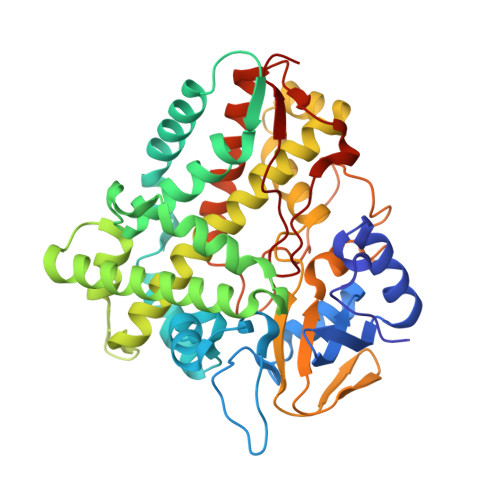
Crystal structures of the ferrous dioxygen complex of wild-type cytochrome P450eryF and its mutants, A245S and A245T: investigation of the proton transfer system in P450eryF.
Nagano, S., Cupp-Vickery, J.R., Poulos, T.L.(2005) J Biol Chem 280: 22102-22107
- PubMed: 15824115
- DOI: https://doi.org/10.1074/jbc.M501732200
- Primary Citation of Related Structures:
1Z8O, 1Z8P, 1Z8Q - PubMed Abstract:
Cytochrome P450eryF (CYP107A) from Saccaropolyspora ertherea catalyzes the hydroxylation of 6-deoxyerythronolide B, one of the early steps in the biosynthesis of erythromycin. P450eryF has an alanine rather than the conserved threonine that participates in the activation of dioxygen (O(2)) in most other P450s. The initial structure of P450eryF (Cupp-Vickery, J. R., Han, O., Hutchinson, C. R., and Poulos, T. L. (1996) Nat. Struct. Biol. 3, 632-637) suggests that the substrate 5-OH replaces the missing threonine OH group and holds a key active site water molecule in position to donate protons to the iron-linked dioxygen, a critical step for the monooxygenase reaction. To probe the proton delivery system in P450eryF, we have solved crystal structures of ferrous wild-type and mutant (Fe(2+)) dioxygen-bound complexes. The catalytic water molecule that was postulated to provide protons to dioxygen is absent, although the substrate 5-OH group donates a hydrogen bond to the iron-linked dioxygen. The hydrogen bond network observed in the wild-type ferrous dioxygen complex, water 63-Glu(360)-Ser(246)-water 53-Ala(241) carbonyl in the I-helix cleft, is proposed as the proton transfer pathway. Consistent with this view, the hydrogen bond network in the O(2).A245S and O(2) .A245T mutants, which have decreased or no enzyme activity, was perturbed or disrupted, respectively. The mutant Thr(245) side chain also perturbs the hydrogen bond between the substrate 5-OH and dioxygen ligand. Contrary to the previously proposed mechanism, these results support the direct involvement of the substrate in O(2) activation but raise questions on the role water plays as a direct proton donor to the iron-linked dioxygen.
Organizational Affiliation:
Departments of Molecular Biology & Biochemistry, Program in Chemical and Structural Biology, University of California-Irvine, Irvine, CA 92697, USA. [email protected]


