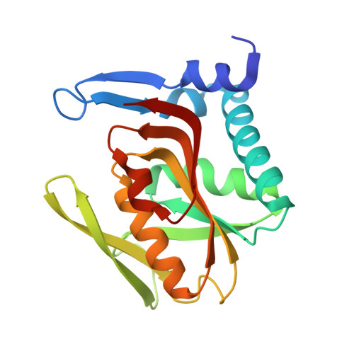
1ZN9

Human Adenine Phosphoribosyltransferase in Apo and AMP Complexed Forms


Experimental Data Snapshot

  • Method: X-RAY DIFFRACTION
  • Resolution: 2.05 Å
  • R-Value Free: 0.225 
  • R-Value Work: 0.151 
  • R-Value Observed: 0.154 

wwPDB Validation   3D Report Full Report


Ligand Structure Quality Assessment 


This is version 1.3 of the entry. See complete history


Literature

Structural Complexes of Human Adenine Phosphoribosyltransferase Reveal Novel Features of the APRT Catalytic Mechanism

Silva, C.H.Silva, M.Iulek, J.Thiemann, O.H.

(2008) J Biomol Struct Dyn 25: 589-598

  • DOI: https://doi.org/10.1080/07391102.2008.10507205
  • Primary Citation of Related Structures:  
    1ZN7, 1ZN8, 1ZN9

  • PubMed Abstract: 

    Adenine phosphoribosyltransferase (APRT) is an important enzyme component of the purine recycling pathway. Parasitic protozoa of the order Kinetoplastida are unable to synthesize purines de novo and use the salvage pathway for the synthesis of purine bases rendering this biosynthetic pathway an attractive target for antiparasitic drug design. The recombinant human adenine phosphoribosyltransferase (hAPRT) structure was resolved in the presence of AMP in the active site to 1.76 A resolution and with the substrates PRPP and adenine simultaneously bound to the catalytic site to 1.83 A resolution. An additional structure was solved containing one subunit of the dimer in the apo-form to 2.10 A resolution. Comparisons of these three hAPRT structures with other 'type I' PRTases revealed several important features of this class of enzymes. Our data indicate that the flexible loop structure adopts an open conformation before and after binding of both substrates adenine and PRPP. Comparative analyses presented here provide structural evidence to propose the role of Glu104 as the residue that abstracts the proton of adenine N9 atom before its nucleophilic attack on the PRPP anomeric carbon. This work leads to new insights to the understanding of the APRT catalytic mechanism.


  • Organizational Affiliation

    Departamento de Física e Informática, Grupo de Cristalografia de Proteínas e Biologia Estrutural, Instituto de Física de São Carlos, USP, Caixa Postal 369, 13560-590, São Carlos-SP, Brazil. [email protected]


Macromolecules
Find similar proteins by:  (by identity cutoff)  |  3D Structure
Entity ID: 1
MoleculeChains Sequence LengthOrganismDetailsImage
Adenine phosphoribosyltransferase
A, B
180Homo sapiensMutation(s): 0 
Gene Names: APRT
EC: 2.4.2.7
UniProt & NIH Common Fund Data Resources
Find proteins for P07741 (Homo sapiens)
Explore P07741 
Go to UniProtKB:  P07741
PHAROS:  P07741
GTEx:  ENSG00000198931 
Entity Groups  
Sequence Clusters30% Identity50% Identity70% Identity90% Identity95% Identity100% Identity
UniProt GroupP07741
Sequence Annotations
Expand
  • Reference Sequence
Small Molecules
Ligands 1 Unique
IDChains Name / Formula / InChI Key2D Diagram3D Interactions
AMP
Query on AMP

Download Ideal Coordinates CCD File 
C [auth B]ADENOSINE MONOPHOSPHATE
C10 H14 N5 O7 P
UDMBCSSLTHHNCD-KQYNXXCUSA-N
Experimental Data & Validation

Experimental Data

  • Method: X-RAY DIFFRACTION
  • Resolution: 2.05 Å
  • R-Value Free: 0.225 
  • R-Value Work: 0.151 
  • R-Value Observed: 0.154 
  • Space Group: P 21 21 21
Unit Cell:
Length ( Å )Angle ( ˚ )
a = 47.511α = 90
b = 49.252β = 90
c = 140.105γ = 90
Software Package:
Software NamePurpose
REFMACrefinement

Structure Validation

View Full Validation Report


Ligand Structure Quality Assessment 


Entry History 

Deposition Data

Revision History  (Full details and data files)

  • Version 1.0: 2006-04-25
    Type: Initial release
  • Version 1.1: 2008-04-21
    Changes: Version format compliance
  • Version 1.2: 2011-07-13
    Changes: Advisory, Version format compliance
  • Version 1.3: 2024-02-14
    Changes: Data collection, Database references, Derived calculations