Proton transfer in the oxidative half-reaction of pentaerythritol tetranitrate reductase
Khan, H., Barna, T., Bruce, N.C., Munro, A.W., Leys, D., Scrutton, N.S.(2005) FEBS J 272: 4660-4671
- PubMed: 16156787
- DOI: https://doi.org/10.1111/j.1742-4658.2005.04875.x
- Primary Citation of Related Structures:
2ABA, 2ABB - PubMed Abstract:
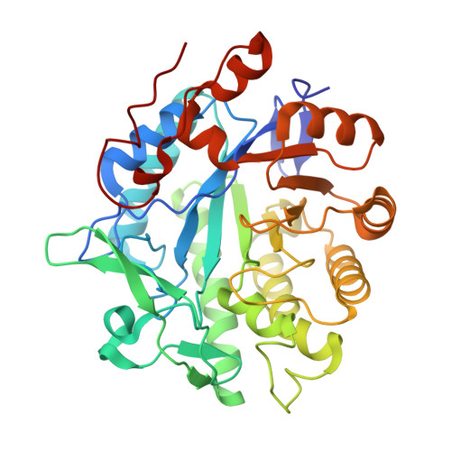
The roles of His181, His184 and Tyr186 in PETN reductase have been examined by mutagenesis, spectroscopic and stopped-flow kinetics, and by determination of crystallographic structures for the Y186F PETN reductase and reduced wild-type enzyme-progesterone complex. Residues His181 and His184 are important in the binding of coenzyme, steroids, nitroaromatic ligands and the substrate 2-cyclohexen-1-one. The H181A and H184A enzymes retain activity in reductive and oxidative half-reactions, and thus do not play an essential role in catalysis. Ligand binding and catalysis is not substantially impaired in Y186F PETN reductase, which contrasts with data for the equivalent mutation (Y196F) in Old Yellow Enzyme. The structure of Y186F PETN reductase is identical to wild-type enzyme, with the obvious exception of the mutation. We show in PETN reductase that Tyr186 is not a key proton donor in the reduction of alpha/beta unsaturated carbonyl compounds. The structure of two electron-reduced PETN reductase bound to the inhibitor progesterone mimics the catalytic enzyme-steroid substrate complex and is similar to the structure of the oxidized enzyme-inhibitor complex. The reactive C1-C2 unsaturated bond of the steroid is inappropriately orientated with the flavin N5 atom for hydride transfer. With steroid substrates, the productive conformation is achieved by orientating the steroid through flipping by 180 degrees , consistent with known geometries for hydride transfer in flavoenzymes. Our data highlight mechanistic differences between Old Yellow Enzyme and PETN reductase and indicate that catalysis requires a metastable enzyme-steroid complex and not the most stable complex observed in crystallographic studies.
Organizational Affiliation:
Department of Biochemistry, University of Leicester, UK.


