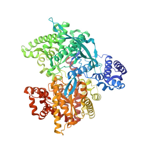
2AW3

X-Ray studies on maltodextrin phosphorylase complexes: recognition of substrates and cathalitic mechanism of phosphorylase family


Experimental Data Snapshot

  • Method: X-RAY DIFFRACTION
  • Resolution: 2.20 Å
  • R-Value Free: 0.236 
  • R-Value Work: 0.175 
  • R-Value Observed: 0.178 

Starting Model: experimental
View more details

wwPDB Validation   3D Report Full Report


Ligand Structure Quality Assessment 


This is version 2.1 of the entry. See complete history


Literature

X-ray studies on ternary complexes of maltodextrin phosphorylase.

Campagnolo, M.Campa, C.Zorzi, R.D.Wuerges, J.Geremia, S.

(2008) Arch Biochem Biophys 471: 11-19

  • DOI: https://doi.org/10.1016/j.abb.2007.11.023
  • Primary Citation of Related Structures:  
    2AV6, 2AW3, 2AZD

  • PubMed Abstract: 

    We report crystal structures of ternary complexes of maltodextrin phosphorylase with natural oligosaccharide and phosphate mimicking anions: nitrate, sulphate and vanadate. Electron density maps obtained from crystals grown in presence of Al(NO3)3 show a nitrate ion instead of the expected AlF4- in the catalytic site. The trigonal NO3- is coplanar with the Arg569 guanidinium group and mimics three of the four oxygen atoms of phosphate. The ternary complex with sulphate shows a partial occupancy of the anionic site. The low affinity of the sulphate ion, observed when the alpha-glucosyl substrate is present in the catalytic channel, is ascribed to restricted space for the anion. Even lower occupancy is observed for the larger vanadate anion. The Malp/G5/VO43- structure shows the partial occupancy of the oligosaccharide and the dislocation of the 380's loop. This has been attributed to the formation of oligosaccharide vanadate derivatives (confirmed by capillary electrophoresis) that reduces their effective concentration. The difficulty to trap a ternary complex mimicking the ground state has been correlated to the apparent lower affinity that natural substrates show regarding the intermediates of the enzymatic reaction.


  • Organizational Affiliation

    CEB-Centre of Excellence in Biocrystallography, Department of Chemical Sciences, University of Trieste, via L. Giorgieri 1, 34127 Trieste, Italy.


Macromolecules
Find similar proteins by:  (by identity cutoff)  |  3D Structure
Entity ID: 1
MoleculeChains Sequence LengthOrganismDetailsImage
Maltodextrin phosphorylase
A, B
796Escherichia coliMutation(s): 0 
Gene Names: EG10560
EC: 2.4.1.1
UniProt
Find proteins for P00490 (Escherichia coli (strain K12))
Explore P00490 
Go to UniProtKB:  P00490
Entity Groups  
Sequence Clusters30% Identity50% Identity70% Identity90% Identity95% Identity100% Identity
UniProt GroupP00490
Sequence Annotations
Expand
  • Reference Sequence
Oligosaccharides

Help

Entity ID: 2
MoleculeChains Length2D Diagram Glycosylation3D Interactions
alpha-D-glucopyranose-(1-4)-alpha-D-glucopyranose-(1-4)-alpha-D-glucopyranose-(1-4)-alpha-D-glucopyranose-(1-4)-beta-D-glucopyranose
C, D
5N/A
Glycosylation Resources
GlyTouCan:  G43441FW
GlyCosmos:  G43441FW
Experimental Data & Validation

Experimental Data

  • Method: X-RAY DIFFRACTION
  • Resolution: 2.20 Å
  • R-Value Free: 0.236 
  • R-Value Work: 0.175 
  • R-Value Observed: 0.178 
  • Space Group: P 21 21 21
Unit Cell:
Length ( Å )Angle ( ˚ )
a = 75.297α = 90
b = 105.887β = 90
c = 219.52γ = 90
Software Package:
Software NamePurpose
REFMACrefinement
MOSFLMdata reduction
CCP4data scaling

Structure Validation

View Full Validation Report


Ligand Structure Quality Assessment 


Entry History 

Deposition Data

Revision History  (Full details and data files)

  • Version 1.0: 2005-09-13
    Type: Initial release
  • Version 1.1: 2008-05-01
    Changes: Version format compliance
  • Version 1.2: 2011-07-13
    Changes: Non-polymer description, Version format compliance
  • Version 2.0: 2020-07-29
    Type: Remediation
    Reason: Carbohydrate remediation
    Changes: Atomic model, Data collection, Derived calculations, Structure summary
  • Version 2.1: 2023-10-25
    Changes: Data collection, Database references, Refinement description, Structure summary