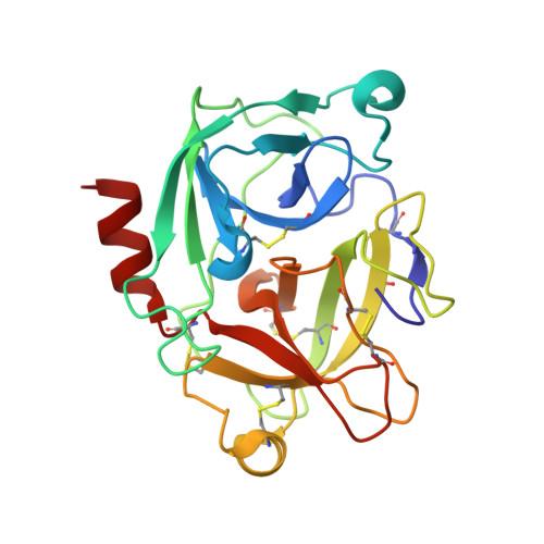
2BDI

Human Kallikrein 4 complex with cobalt and p-aminobenzamidine


Experimental Data Snapshot

  • Method: X-RAY DIFFRACTION
  • Resolution: 3.00 Å
  • R-Value Free: 0.296 
  • R-Value Work: 0.258 
  • R-Value Observed: 0.258 

Starting Model: other
View more details

wwPDB Validation   3D Report Full Report


This is version 1.4 of the entry. See complete history


Literature

Crystal structures of human tissue kallikrein 4: activity modulation by a specific zinc binding site.

Debela, M.Magdolen, V.Grimminger, V.Sommerhoff, C.Messerschmidt, A.Huber, R.Friedrich, R.Bode, W.Goettig, P.

(2006) J Mol Biol 362: 1094-1107

  • DOI: https://doi.org/10.1016/j.jmb.2006.08.003
  • Primary Citation of Related Structures:  
    2BDG, 2BDH, 2BDI

  • PubMed Abstract: 

    Human tissue kallikrein 4 (hK4) belongs to a 15-member family of closely related serine proteinases. hK4 is predominantly expressed in prostate, activates hK3/PSA, and is up-regulated in prostate and ovarian cancer. We have identified active monomers of recombinant hK4 besides inactive oligomers in solution. hK4 crystallised in the presence of zinc, nickel, and cobalt ions in three crystal forms containing cyclic tetramers and octamers. These structures display a novel metal site between His25 and Glu77 that links the 70-80 loop with the N-terminal segment. Micromolar zinc as present in prostatic fluid inhibits the enzymatic activity of hK4 against fluorogenic substrates. In our measurements, wild-type hK4 exhibited a zinc inhibition constant (IC50) of 16 microM including a permanent residual activity, in contrast to the zinc-independent mutants H25A and E77A. Since the Ile16 N terminus of wild-type hK4 becomes more accessible for acetylating agents in the presence of zinc, we propose that zinc affects the hK4 active site via the salt-bridge formed between the N terminus and Asp194 required for a functional active site. hK4 possesses an unusual 99-loop that creates a groove-like acidic S2 subsite. These findings explain the observed specificity of hK4 for the P1 to P4 substrate residues. Moreover, hK4 shows a negatively charged surface patch, which may represent an exosite for prime-side substrate recognition.


  • Organizational Affiliation

    Max-Planck-Institut für Biochemie, Proteinase Research Group, Am Klopferspitz 18, 82152 Martinsried, Germany.


Macromolecules
Find similar proteins by:  (by identity cutoff)  |  3D Structure
Entity ID: 1
MoleculeChains Sequence LengthOrganismDetailsImage
Kallikrein-4
A, B, C, D, E
A, B, C, D, E, F, G, H, I, J, K, L, M, N, O, P
223Homo sapiensMutation(s): 0 
Gene Names: KLK4EMSP1PRSS17PSTS
EC: 3.4.21
UniProt & NIH Common Fund Data Resources
Find proteins for Q9Y5K2 (Homo sapiens)
Explore Q9Y5K2 
Go to UniProtKB:  Q9Y5K2
PHAROS:  Q9Y5K2
GTEx:  ENSG00000167749 
Entity Groups  
Sequence Clusters30% Identity50% Identity70% Identity90% Identity95% Identity100% Identity
UniProt GroupQ9Y5K2
Sequence Annotations
Expand
  • Reference Sequence
Small Molecules
Ligands 2 Unique
IDChains Name / Formula / InChI Key2D Diagram3D Interactions
PBZ
Query on PBZ

Download Ideal Coordinates CCD File 
BA [auth I]
CA [auth J]
DA [auth K]
EA [auth L]
GA [auth M]
BA [auth I],
CA [auth J],
DA [auth K],
EA [auth L],
GA [auth M],
HA [auth N],
IA [auth O],
JA [auth P],
R [auth A],
S [auth B],
T [auth C],
U [auth D],
W [auth E],
X [auth F],
Y [auth G],
Z [auth H]
P-AMINO BENZAMIDINE
C7 H10 N3
WPANETAWYGDRLL-UHFFFAOYSA-O
CO
Query on CO

Download Ideal Coordinates CCD File 
AA [auth I],
FA [auth M],
Q [auth A],
V [auth E]
COBALT (II) ION
Co
XLJKHNWPARRRJB-UHFFFAOYSA-N
Experimental Data & Validation

Experimental Data

  • Method: X-RAY DIFFRACTION
  • Resolution: 3.00 Å
  • R-Value Free: 0.296 
  • R-Value Work: 0.258 
  • R-Value Observed: 0.258 
  • Space Group: P 1 21 1
Unit Cell:
Length ( Å )Angle ( ˚ )
a = 147.05α = 90
b = 73.86β = 102.41
c = 154.38γ = 90
Software Package:
Software NamePurpose
CNSrefinement
DENZOdata reduction
SCALEPACKdata scaling
PHASERphasing

Structure Validation

View Full Validation Report


Entry History 

Deposition Data

Revision History  (Full details and data files)

  • Version 1.0: 2006-10-03
    Type: Initial release
  • Version 1.1: 2008-05-01
    Changes: Version format compliance
  • Version 1.2: 2011-07-13
    Changes: Version format compliance
  • Version 1.3: 2024-04-03
    Changes: Data collection, Database references, Derived calculations, Refinement description
  • Version 1.4: 2024-10-30
    Changes: Structure summary