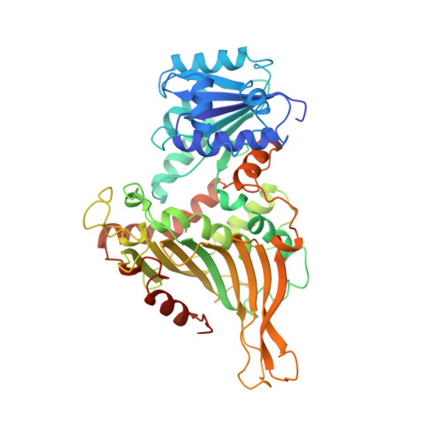
2BHL

X-RAY STRUCTURE OF HUMAN GLUCOSE-6-PHOSPHATE DEHYDROGENASE (DELETION VARIANT) COMPLEXED WITH GLUCOSE-6-PHOSPHATE


Experimental Data Snapshot

  • Method: X-RAY DIFFRACTION
  • Resolution: 2.90 Å
  • R-Value Free: 0.261 
  • R-Value Work: 0.212 
  • R-Value Observed: 0.214 

Starting Model: experimental
View more details

wwPDB Validation   3D Report Full Report


Ligand Structure Quality Assessment 


This is version 1.4 of the entry. See complete history


Literature

Structural Studies of Glucose-6-Phosphate and Nadp+ Binding to Human Glucose-6-Phosphate Dehydrogenase

Kotaka, M.Gover, S.Vandeputte-Rutten, L.Au, S.W.N.Lam, V.M.S.Adams, M.J.

(2005) Acta Crystallogr D Biol Crystallogr 61: 495

  • DOI: https://doi.org/10.1107/S0907444905002350
  • Primary Citation of Related Structures:  
    2BH9, 2BHL

  • PubMed Abstract: 

    Human glucose-6-phosphate dehydrogenase (G6PD) is NADP(+)-dependent and catalyses the first and rate-limiting step of the pentose phosphate shunt. Binary complexes of the human deletion mutant, DeltaG6PD, with glucose-6-phosphate and NADP(+) have been crystallized and their structures solved to 2.9 and 2.5 A, respectively. The structures are compared with the previously determined structure of the Canton variant of human G6PD (G6PD(Canton)) in which NADP(+) is bound at the structural site. Substrate binding in DeltaG6PD is shown to be very similar to that described previously in Leuconostoc mesenteroides G6PD. NADP(+) binding at the coenzyme site is seen to be comparable to NADP(+) binding in L. mesenteroides G6PD, although some differences arise as a result of sequence changes. The tetramer interface varies slightly among the human G6PD complexes, suggesting flexibility in the predominantly hydrophilic dimer-dimer interactions. In both complexes, Pro172 of the conserved peptide EKPxG is in the cis conformation; it is seen to be crucial for close approach of the substrate and coenzyme during the enzymatic reaction. Structural NADP(+) binds in a very similar way in the DeltaG6PD-NADP(+) complex and in G6PD(Canton), while in the substrate complex the structural NADP(+) has low occupancy and the C-terminal tail at the structural NADP(+) site is disordered. The implications of possible interaction between the structural NADP(+) and G6P are considered.


  • Organizational Affiliation

    Laboratory of Molecular Biophysics, Department of Biochemistry, University of Oxford, South Parks Road, Oxford OX1 3QU, England. [email protected]


Macromolecules
Find similar proteins by:  (by identity cutoff)  |  3D Structure
Entity ID: 1
MoleculeChains Sequence LengthOrganismDetailsImage
GLUCOSE-6-PHOSPHATE 1-DEHYDROGENASE
A, B
489Homo sapiensMutation(s): 0 
EC: 1.1.1.49
UniProt & NIH Common Fund Data Resources
Find proteins for P11413 (Homo sapiens)
Explore P11413 
Go to UniProtKB:  P11413
PHAROS:  P11413
GTEx:  ENSG00000160211 
Entity Groups  
Sequence Clusters30% Identity50% Identity70% Identity90% Identity95% Identity100% Identity
UniProt GroupP11413
Sequence Annotations
Expand
  • Reference Sequence
Small Molecules
Ligands 2 Unique
IDChains Name / Formula / InChI Key2D Diagram3D Interactions
BG6
Query on BG6

Download Ideal Coordinates CCD File 
G [auth A],
J [auth B]
6-O-phosphono-beta-D-glucopyranose
C6 H13 O9 P
NBSCHQHZLSJFNQ-VFUOTHLCSA-N
GOL
Query on GOL

Download Ideal Coordinates CCD File 
C [auth A]
D [auth A]
E [auth A]
F [auth A]
H [auth B]
C [auth A],
D [auth A],
E [auth A],
F [auth A],
H [auth B],
I [auth B]
GLYCEROL
C3 H8 O3
PEDCQBHIVMGVHV-UHFFFAOYSA-N
Experimental Data & Validation

Experimental Data

  • Method: X-RAY DIFFRACTION
  • Resolution: 2.90 Å
  • R-Value Free: 0.261 
  • R-Value Work: 0.212 
  • R-Value Observed: 0.214 
  • Space Group: C 2 2 21
Unit Cell:
Length ( Å )Angle ( ˚ )
a = 117.552α = 90
b = 179.527β = 90
c = 137.938γ = 90
Software Package:
Software NamePurpose
TNTrefinement
MOSFLMdata reduction
SCALAdata scaling
MOLREPphasing

Structure Validation

View Full Validation Report


Ligand Structure Quality Assessment 


Entry History 

Deposition Data

Revision History  (Full details and data files)

  • Version 1.0: 2005-04-25
    Type: Initial release
  • Version 1.1: 2014-02-05
    Changes: Other, Refinement description, Structure summary, Version format compliance
  • Version 1.2: 2019-05-22
    Changes: Data collection, Other, Refinement description
  • Version 1.3: 2020-07-29
    Type: Remediation
    Reason: Carbohydrate remediation
    Changes: Data collection, Derived calculations, Other, Structure summary
  • Version 1.4: 2023-12-13
    Changes: Data collection, Database references, Refinement description, Structure summary