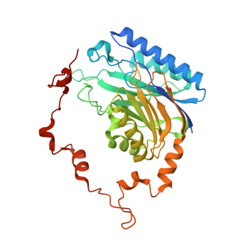
2E2L

Helicobacter pylori formamidase AmiF contains a fine-tuned cysteine-glutamate-lysine catalytic triad


Experimental Data Snapshot

  • Method: X-RAY DIFFRACTION
  • Resolution: 2.29 Å
  • R-Value Free: 0.285 
  • R-Value Work: 0.253 
  • R-Value Observed: 0.255 

Starting Model: experimental
View more details

wwPDB Validation   3D Report Full Report


This is version 1.4 of the entry. See complete history


Literature

Crystal structure of Helicobacter pylori formamidase AmiF reveals a cysteine-glutamate-lysine catalytic triad

Hung, C.-L.Liu, J.-H.Chiu, W.-C.Huang, S.-W.Hwang, J.-K.Wang, W.-C.

(2007) J Biol Chem 282: 12220-12229

  • DOI: https://doi.org/10.1074/jbc.M609134200
  • Primary Citation of Related Structures:  
    2DYU, 2DYV, 2E2K, 2E2L

  • PubMed Abstract: 

    Helicobacter pylori AmiF formamidase that hydrolyzes formamide to produce formic acid and ammonia belongs to a member of the nitrilase superfamily. The crystal structure of AmiF was solved to 1.75A resolution using single-wavelength anomalous dispersion methods. The structure consists of a homohexamer related by 3-fold symmetry in which each subunit has an alpha-beta-beta-alpha four-layer architecture characteristic of the nitrilase superfamily. One exterior alpha layer faces the solvent, whereas the other one associates with that of the neighbor subunit, forming a tight alpha-beta-beta-alpha-alpha-beta-beta-alpha dimer. The apo and liganded crystal structures of an inactive mutant C166S were also determined to 2.50 and 2.30 A, respectively. These structures reveal a small formamide-binding pocket that includes Cys(166), Glu(60), and Lys(133) catalytic residues, in which Cys(166) acts as a nucleophile. Analysis of the liganded AmiF and N-carbamoyl d-amino acid amidohydrolase binding pockets reveals a common Cys-Glu-Lys triad, another conserved glutamate, and different subsets of ligand-binding residues. Molecular dynamic simulations show that the conserved triad has minimal fluctuations, catalyzing the hydrolysis of a specific nitrile or amide in the nitrilase superfamily efficiently.


  • Organizational Affiliation

    Institute of Molecular and Cellular Biology and Department of Life Science, National Tsing Hua University, Hsinchu 300, Taiwan.


Macromolecules
Find similar proteins by:  (by identity cutoff)  |  3D Structure
Entity ID: 1
MoleculeChains Sequence LengthOrganismDetailsImage
Formamidase
A, B, C, D, E
A, B, C, D, E, F
334Helicobacter pylori 26695Mutation(s): 1 
Gene Names: amiF
EC: 3.5.1.49
UniProt
Find proteins for O25836 (Helicobacter pylori (strain ATCC 700392 / 26695))
Explore O25836 
Go to UniProtKB:  O25836
Entity Groups  
Sequence Clusters30% Identity50% Identity70% Identity90% Identity95% Identity100% Identity
UniProt GroupO25836
Sequence Annotations
Expand
  • Reference Sequence
Experimental Data & Validation

Experimental Data

  • Method: X-RAY DIFFRACTION
  • Resolution: 2.29 Å
  • R-Value Free: 0.285 
  • R-Value Work: 0.253 
  • R-Value Observed: 0.255 
  • Space Group: P 1 21 1
Unit Cell:
Length ( Å )Angle ( ˚ )
a = 83.089α = 90
b = 151.795β = 114.99
c = 89.083γ = 90
Software Package:
Software NamePurpose
REFMACrefinement
HKL-2000data collection
HKL-2000data reduction
HKL-2000data scaling
AMoREphasing

Structure Validation

View Full Validation Report


Entry History 

Deposition Data

Revision History  (Full details and data files)

  • Version 1.0: 2007-02-13
    Type: Initial release
  • Version 1.1: 2008-04-30
    Changes: Version format compliance
  • Version 1.2: 2011-07-13
    Changes: Advisory, Source and taxonomy, Version format compliance
  • Version 1.3: 2021-11-10
    Changes: Database references, Derived calculations
  • Version 1.4: 2023-10-25
    Changes: Data collection, Refinement description