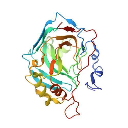
Carbonic Anhydrase Activators. Activation of Isozymes I, II, IV, VA, VII, and XIV with L- and D-Histidine and Crystallographic Analysis of Their Adducts with Isoform II: Engineering Proton-Transfer Processes within the Active Site of an Enzyme.
Temperini, C., Scozzafava, A., Vullo, D., Supuran, C.T.(2006) Chemistry 12: 7057-7066
- PubMed: 16807956
- DOI: https://doi.org/10.1002/chem.200600159
- Primary Citation of Related Structures:
2EZ7 - PubMed Abstract:
Activation of six human carbonic anhydrases (CA, EC 4.2.1.1), that is, hCA I, II, IV, VA, VII, and XIV, with l- and d-histidine was investigated through kinetics and by X-ray crystallography. l-His was a potent activator of isozymes I, VA, VII, and XIV, and a weaker activator of hCA II and IV. d-His showed good hCA I, VA, and VII activation properties, being a moderate activator of hCA XIV and a weak activator of hCA II and IV. The structures as determined by X-ray crystallography of the hCA II-l-His/d-His adducts showed the activators to be anchored at the entrance of the active site, contributing to extended networks of hydrogen bonds with amino acid residues/water molecules present in the cavity, explaining their different potency and interaction patterns with various isozymes. The residues involved in l-His recognition were His64, Asn67, Gln92, whereas three water molecules connected the activator to the zinc-bound hydroxide. Only the imidazole moiety of l-His interacted with these amino acids. For the d-His adduct, the residues involved in recognition of the activator were Trp5, His64, and Pro201, whereas two water molecules connected the zinc-bound water to the activator. Only the COOH and NH(2) moieties of d-His participated in hydrogen bonds with these residues. This is the first study showing different binding modes of stereoisomeric activators within the hCA II active site, with consequences for overall proton-transfer processes (rate-determining for the catalytic cycle). The study also points out differences of activation efficiency between various isozymes with structurally related activators, convenient for designing alternative proton-transfer pathways, useful both for a better understanding of the catalytic mechanism and for obtaining pharmacologically useful derivatives, for example, for the management of Alzheimer's disease.
Organizational Affiliation:
Università degli Studi di Firenze, Laboratorio di Chimica Bioinorganica, Via della Lastruccia 3, I-50019 Sesto Fiorentino (Firenze), Italy.


