2EZT

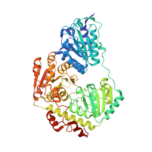
Pyruvate oxidase variant F479W in complex with reaction intermediate 2-hydroxyethyl-thiamin diphosphate


Experimental Data Snapshot

  • Method: X-RAY DIFFRACTION
  • Resolution: 2.29 Å
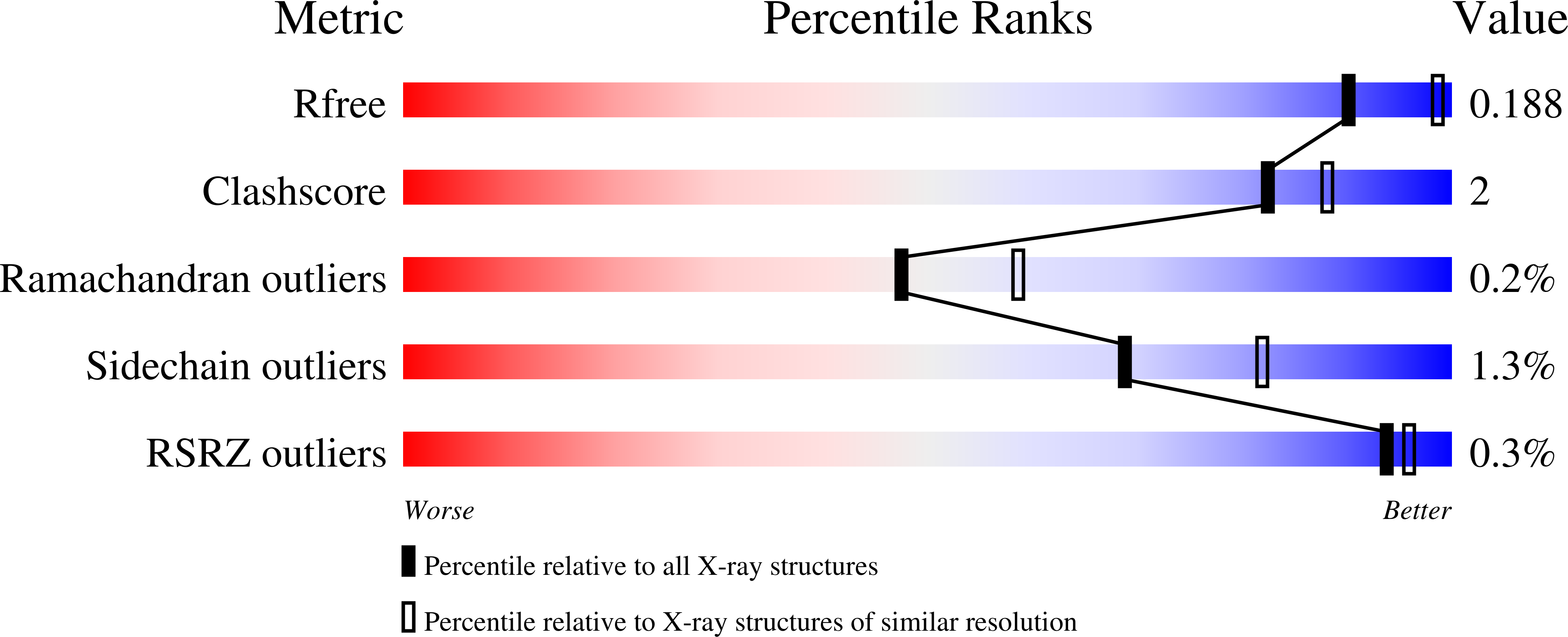
  • R-Value Free: 0.187 
  • R-Value Work: 0.150 
  • R-Value Observed: 0.151 

Starting Model: experimental
View more details

wwPDB Validation   3D Report Full Report


Ligand Structure Quality Assessment 


This is version 2.0 of the entry. See complete history


Literature

The catalytic cycle of a thiamin diphosphate enzyme examined by cryocrystallography.

Wille, G.Meyer, D.Steinmetz, A.Hinze, E.Golbik, R.Tittmann, K.

(2006) Nat Chem Biol 2: 324-328

  • DOI: https://doi.org/10.1038/nchembio788
  • Primary Citation of Related Structures:  
    2EZ4, 2EZ8, 2EZ9, 2EZT, 2EZU

  • PubMed Abstract: 

    Enzymes that use the cofactor thiamin diphosphate (ThDP, 1), the biologically active form of vitamin B(1), are involved in numerous metabolic pathways in all organisms. Although a theory of the cofactor's underlying reaction mechanism has been established over the last five decades, the three-dimensional structures of most major reaction intermediates of ThDP enzymes have remained elusive. Here, we report the X-ray structures of key intermediates in the oxidative decarboxylation of pyruvate, a central reaction in carbon metabolism catalyzed by the ThDP- and flavin-dependent enzyme pyruvate oxidase (POX)3 from Lactobacillus plantarum. The structures of 2-lactyl-ThDP (LThDP, 2) and its stable phosphonate analog, of 2-hydroxyethyl-ThDP (HEThDP, 3) enamine and of 2-acetyl-ThDP (AcThDP, 4; all shown bound to the enzyme's active site) provide profound insights into the chemical mechanisms and the stereochemical course of thiamin catalysis. These snapshots also suggest a mechanism for a phosphate-linked acyl transfer coupled to electron transfer in a radical reaction of pyruvate oxidase.


  • Organizational Affiliation

    Institut für Biochemie, Martin-Luther-Universität Halle-Wittenberg, Kurt-Mothes-Str. 3, 06120 Halle/Saale, Germany. [email protected]


Macromolecules
Find similar proteins by:  (by identity cutoff)  |  3D Structure
Entity ID: 1
MoleculeChains Sequence LengthOrganismDetailsImage
Pyruvate oxidase
A, B
603Lactiplantibacillus plantarumMutation(s): 1 
Gene Names: pox5
EC: 1.2.3.3
UniProt
Find proteins for P37063 (Lactiplantibacillus plantarum (strain ATCC BAA-793 / NCIMB 8826 / WCFS1))
Explore P37063 
Go to UniProtKB:  P37063
Entity Groups  
Sequence Clusters30% Identity50% Identity70% Identity90% Identity95% Identity100% Identity
UniProt GroupP37063
Sequence Annotations
Expand
  • Reference Sequence
Small Molecules
Ligands 6 Unique
IDChains Name / Formula / InChI Key2D Diagram3D Interactions
FAD
Query on FAD

Download Ideal Coordinates CCD File 
G [auth A],
M [auth B]
FLAVIN-ADENINE DINUCLEOTIDE
C27 H33 N9 O15 P2
VWWQXMAJTJZDQX-UYBVJOGSSA-N
TDM
Query on TDM

Download Ideal Coordinates CCD File 
F [auth A],
L [auth B]
2-[(2E)-3-[(4-AMINO-2-METHYLPYRIMIDIN-5-YL)METHYL]-2-(1-HYDROXYETHYLIDENE)-4-METHYL-2,3-DIHYDRO-1,3-THIAZOL-5-YL]ETHYL TRIHYDROGEN DIPHOSPHATE
C14 H22 N4 O8 P2 S
ZZLCKFKIIVXBQU-NTEUORMPSA-N
PO4
Query on PO4

Download Ideal Coordinates CCD File 
D [auth A],
K [auth B]
PHOSPHATE ION
O4 P
NBIIXXVUZAFLBC-UHFFFAOYSA-K
PYR
Query on PYR

Download Ideal Coordinates CCD File 
H [auth A],
N [auth B]
PYRUVIC ACID
C3 H4 O3
LCTONWCANYUPML-UHFFFAOYSA-N
MG
Query on MG

Download Ideal Coordinates CCD File 
C [auth A],
J [auth B]
MAGNESIUM ION
Mg
JLVVSXFLKOJNIY-UHFFFAOYSA-N
NA
Query on NA

Download Ideal Coordinates CCD File 
E [auth A],
I [auth B]
SODIUM ION
Na
FKNQFGJONOIPTF-UHFFFAOYSA-N
Experimental Data & Validation

Experimental Data

  • Method: X-RAY DIFFRACTION
  • Resolution: 2.29 Å
  • R-Value Free: 0.187 
  • R-Value Work: 0.150 
  • R-Value Observed: 0.151 
  • Space Group: C 2 2 21
Unit Cell:
Length ( Å )Angle ( ˚ )
a = 118.79α = 90
b = 154.022β = 90
c = 165.32γ = 90
Software Package:
Software NamePurpose
DENZOdata reduction
SCALEPACKdata scaling
REFMACrefinement
PDB_EXTRACTdata extraction
MOLREPphasing

Structure Validation

View Full Validation Report


Ligand Structure Quality Assessment 


Entry History 

Deposition Data

Revision History  (Full details and data files)

  • Version 1.0: 2006-04-25
    Type: Initial release
  • Version 1.1: 2008-05-01
    Changes: Version format compliance
  • Version 1.2: 2011-07-13
    Changes: Derived calculations, Version format compliance
  • Version 1.3: 2017-10-18
    Changes: Refinement description
  • Version 1.4: 2021-10-20
    Changes: Data collection, Database references, Derived calculations
  • Version 1.5: 2023-08-23
    Changes: Data collection, Refinement description
  • Version 2.0: 2023-11-15
    Changes: Atomic model, Data collection