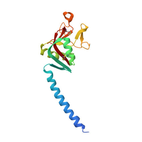
2GGX

Crystal structure of the trimer neck and carbohydrate recognition domain of human surfactant protein D in complex with p-nitrophenyl maltoside


Experimental Data Snapshot

  • Method: X-RAY DIFFRACTION
  • Resolution: 1.90 Å
  • R-Value Free: 0.238 
  • R-Value Work: 0.214 
  • R-Value Observed: 0.220 

Starting Model: experimental
View more details

wwPDB Validation   3D Report Full Report


Ligand Structure Quality Assessment 


This is version 1.5 of the entry. See complete history


Literature

Contributions of Phenylalanine 335 to Ligand Recognition by Human Surfactant Protein D: ring interactions with Sp-D ligands

Crouch, E.McDonald, B.Smith, K.Cafarella, T.Seaton, B.Head, J.

(2006) J Biol Chem 281: 18008-18014

  • DOI: https://doi.org/10.1074/jbc.M601749200
  • Primary Citation of Related Structures:  
    2GGU, 2GGX

  • PubMed Abstract: 

    Surfactant protein D (SP-D) is an innate immune effector that contributes to antimicrobial host defense and immune regulation. Interactions of SP-D with microorganisms and organic antigens involve binding of glycoconjugates to the C-type lectin carbohydrate recognition domain (CRD). A trimeric fusion protein encoding the human neck+CRD bound to the aromatic glycoside p-nitrophenyl-alpha-D-maltoside with nearly a log-fold higher affinity than maltose, the prototypical competitor. Maltotriose, which has the same linkage pattern as the maltoside, bound with intermediate affinity. Site-directed substitution of leucine for phenylalanine 335 (Phe-335) decreased affinities for the maltoside and maltotriose without significantly altering the affinity for maltose or glucose, and substitution of tyrosine or tryptophan for leucine restored preferential binding to maltotriose and the maltoside. A mutant with alanine at this position failed to bind to mannan or maltose-substituted solid supports. Crystallographic analysis of the human neck+CRD complexed with maltotriose or p-nitrophenyl-maltoside showed stacking of the terminal glucose or nitrophenyl ring with the aromatic ring of Phe-335. Our studies indicate that Phe-335, which is evolutionarily conserved in all known SP-Ds, plays important, if not critical, roles in SP-D function.


  • Organizational Affiliation

    Department of Pathology and Immunology, Washington University School of Medicine, 660 S. Euclid Avenue, St. Louis, MO 63110, USA. [email protected]


Macromolecules
Find similar proteins by:  (by identity cutoff)  |  3D Structure
Entity ID: 1
MoleculeChains Sequence LengthOrganismDetailsImage
Pulmonary surfactant-associated protein D
A, B, C
160Homo sapiensMutation(s): 0 
Gene Names: SFTPD
UniProt & NIH Common Fund Data Resources
Find proteins for P35247 (Homo sapiens)
Explore P35247 
Go to UniProtKB:  P35247
PHAROS:  P35247
GTEx:  ENSG00000133661 
Entity Groups  
Sequence Clusters30% Identity50% Identity70% Identity90% Identity95% Identity100% Identity
UniProt GroupP35247
Sequence Annotations
Expand
  • Reference Sequence
Small Molecules
Ligands 2 Unique
IDChains Name / Formula / InChI Key2D Diagram3D Interactions
NPJ
Query on NPJ

Download Ideal Coordinates CCD File 
G [auth A],
K [auth B],
O [auth C]
4-NITROPHENYL 4-O-ALPHA-D-GLUCOPYRANOSYL-ALPHA-D-GALACTOPYRANOSIDE
C18 H25 N O13
IAYJZWFYUSNIPN-LTHBGAKLSA-N
CA
Query on CA

Download Ideal Coordinates CCD File 
D [auth A]
E [auth A]
F [auth A]
H [auth B]
I [auth B]
D [auth A],
E [auth A],
F [auth A],
H [auth B],
I [auth B],
J [auth B],
L [auth C],
M [auth C],
N [auth C]
CALCIUM ION
Ca
BHPQYMZQTOCNFJ-UHFFFAOYSA-N
Binding Affinity Annotations 
IDSourceBinding Affinity
NPJ PDBBind:  2GGX IC50: 3.20e+5 (nM) from 1 assay(s)
Experimental Data & Validation

Experimental Data

  • Method: X-RAY DIFFRACTION
  • Resolution: 1.90 Å
  • R-Value Free: 0.238 
  • R-Value Work: 0.214 
  • R-Value Observed: 0.220 
  • Space Group: P 1 21 1
Unit Cell:
Length ( Å )Angle ( ˚ )
a = 55.431α = 90
b = 106.4β = 92.65
c = 55.08γ = 90
Software Package:
Software NamePurpose
DENZOdata reduction
SCALEPACKdata scaling
CNSrefinement
PDB_EXTRACTdata extraction
ADSCdata collection
CNSphasing

Structure Validation

View Full Validation Report


Ligand Structure Quality Assessment 


Entry History 

Deposition Data

  • Released Date: 2006-05-02 
  • Deposition Author(s): Head, J.

Revision History  (Full details and data files)

  • Version 1.0: 2006-05-02
    Type: Initial release
  • Version 1.1: 2008-05-01
    Changes: Version format compliance
  • Version 1.2: 2011-07-13
    Changes: Version format compliance
  • Version 1.3: 2017-10-18
    Changes: Refinement description
  • Version 1.4: 2023-08-30
    Changes: Data collection, Database references, Derived calculations, Refinement description
  • Version 1.5: 2024-10-30
    Changes: Structure summary