2H4X

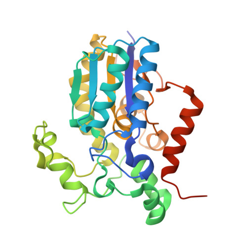
Human bisphosphoglycerate mutase complex with 3-phosphoglycerate with crystal growth 90 days


Experimental Data Snapshot

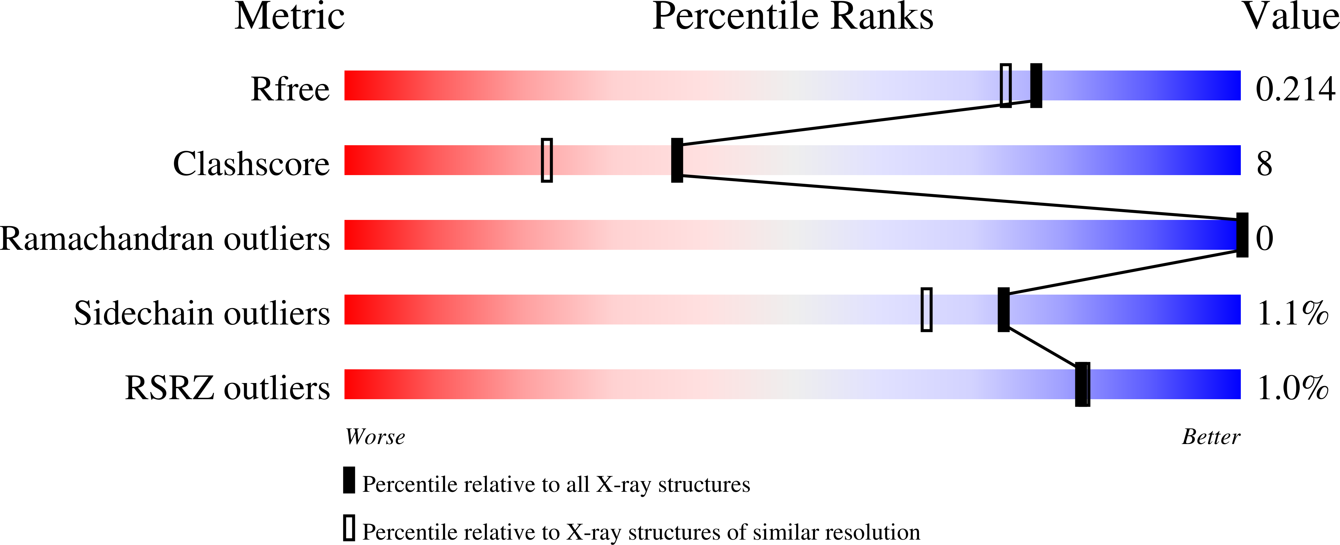
  • Method: X-RAY DIFFRACTION
  • Resolution: 1.85 Å
  • R-Value Free: 0.215 
  • R-Value Work: 0.180 
  • R-Value Observed: 0.180 

Starting Model: experimental
View more details

wwPDB Validation   3D Report Full Report


Ligand Structure Quality Assessment 


This is version 1.3 of the entry. See complete history


Literature

Seeing the process of histidine phosphorylation in human bisphosphoglycerate mutase.

Wang, Y.Liu, L.Wei, Z.Cheng, Z.Lin, Y.Gong, W.

(2006) J Biol Chem 281: 39642-39648

  • DOI: https://doi.org/10.1074/jbc.M606421200
  • Primary Citation of Related Structures:  
    2A9J, 2F90, 2H4X, 2H4Z, 2HHJ

  • PubMed Abstract: 

    Bisphosphoglycerate mutase is an erythrocyte-specific enzyme catalyzing a series of intermolecular phosphoryl group transfer reactions. Its main function is to synthesize 2,3-bisphosphoglycerate, the allosteric effector of hemoglobin. In this paper, we directly observed real-time motion of the enzyme active site and the substrate during phosphoryl transfer. A series of high resolution crystal structures of human bisphosphoglycerate mutase co-crystallized with 2,3-bisphosphoglycerate, representing different time points in the phosphoryl transfer reaction, were solved. These structures not only clarify the argument concerning the substrate binding mode for this enzyme family but also depict the entire process of the key histidine phosphorylation as a "slow movie". It was observed that the enzyme conformation continuously changed during the different states of the reaction. These results provide direct evidence for an "in line" phosphoryl transfer mechanism, and the roles of some key residues in the phosphoryl transfer process are identified.


  • Organizational Affiliation

    National Laboratory of Biomacromolecules, Institute of Biophysics, Chinese Academy of Sciences, Beijing 100101, China.


Macromolecules
Find similar proteins by:  (by identity cutoff)  |  3D Structure
Entity ID: 1
MoleculeChains Sequence LengthOrganismDetailsImage
Bisphosphoglycerate mutase
A, B
267Homo sapiensMutation(s): 0 
EC: 5.4.2.4 (PDB Primary Data), 5.4.2.1 (PDB Primary Data), 3.1.3.13 (PDB Primary Data), 5.4.2.11 (UniProt)
UniProt & NIH Common Fund Data Resources
Find proteins for P07738 (Homo sapiens)
Explore P07738 
Go to UniProtKB:  P07738
PHAROS:  P07738
GTEx:  ENSG00000172331 
Entity Groups  
Sequence Clusters30% Identity50% Identity70% Identity90% Identity95% Identity100% Identity
UniProt GroupP07738
Sequence Annotations
Expand
  • Reference Sequence
Experimental Data & Validation

Experimental Data

  • Method: X-RAY DIFFRACTION
  • Resolution: 1.85 Å
  • R-Value Free: 0.215 
  • R-Value Work: 0.180 
  • R-Value Observed: 0.180 
  • Space Group: P 21 21 21
Unit Cell:
Length ( Å )Angle ( ˚ )
a = 48.498α = 90
b = 71.362β = 90
c = 160.162γ = 90
Software Package:
Software NamePurpose
DENZOdata reduction
SCALEPACKdata scaling
AMoREphasing
CNSrefinement

Structure Validation

View Full Validation Report


Ligand Structure Quality Assessment 


Entry History 

Deposition Data

Revision History  (Full details and data files)

  • Version 1.0: 2006-10-24
    Type: Initial release
  • Version 1.1: 2008-05-01
    Changes: Version format compliance
  • Version 1.2: 2011-07-13
    Changes: Version format compliance
  • Version 1.3: 2023-10-25
    Changes: Data collection, Database references, Derived calculations, Refinement description