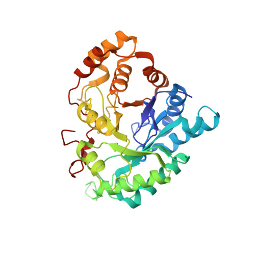
2IPJ

Crystal structure of h3alpha-hydroxysteroid dehydrogenase type 3 mutant Y24A in complex with NADP+ and epi-testosterone


Experimental Data Snapshot

  • Method: X-RAY DIFFRACTION
  • Resolution: 1.80 Å
  • R-Value Free: 0.228 
  • R-Value Work: 0.204 

Starting Model: experimental
View more details

wwPDB Validation   3D Report Full Report


Ligand Structure Quality Assessment 


This is version 1.5 of the entry. See complete history


Literature

Mouse 17alpha-Hydroxysteroid Dehydrogenase (AKR1C21) Binds Steroids Differently from other Aldo-keto Reductases: Identification and Characterization of Amino Acid Residues Critical for Substrate Binding.

Faucher, F.Cantin, L.Pereira de Jesus-Tran, K.Lemieux, M.Luu-The, V.Labrie, F.Breton, R.

(2007) J Mol Biol 369: 525-540

  • DOI: https://doi.org/10.1016/j.jmb.2007.03.058
  • Primary Citation of Related Structures:  
    2IPF, 2IPG, 2IPJ

  • PubMed Abstract: 

    The mouse 17alpha-hydroxysteroid dehydrogenase (m17alpha-HSD) is the unique known member of the aldo-keto reductase (AKR) superfamily able to catalyze efficiently and in a stereospecific manner the conversion of androstenedione (Delta4) into epi-testosterone (epi-T), the 17alpha-epimer of testosterone. Structural and mutagenic studies had already identified one of the residues delineating the steroid-binding cavity, A24, as the major molecular determinant for the stereospecificity of m17alpha-HSD. We report here a ternary complex crystal structure (m17alpha-HSD:NADP(+):epi-T) determined at 1.85 A resolution that confirms this and reveals a unique steroid-binding mode for an AKR enzyme. Indeed, in addition to the interactions found in all other AKRs (van der Waals contacts stabilizing the core of the steroid and the hydrogen bonds established at the catalytic site by the Y55 and H117 residues with the oxygen atom of the ketone group to be reduced), m17alpha-HSD establishes with the other extremity of the steroid nucleus an additional interaction involving K31. By combining direct mutagenesis and kinetic studies, we found that the elimination of this hydrogen bond did not affect the affinity of the enzyme for its steroid substrate but led to a slight but significant increase of its catalytic efficiency (k(cat)/K(m)), suggesting a role for K31 in the release of the steroidal product at the end of the reaction. This previously unobserved steroid-binding mode for an AKR is similar to that adopted by other steroid-binding proteins, the hydroxysteroid dehydrogenases of the short-chain dehydrogenases/reductases (SDR) family and the steroid hormone nuclear receptors. Mutagenesis and structural studies made on the human type 3 3alpha-HSD, a closely related enzyme that shares 73% amino acids identity with the m17alpha-HSD, also revealed that the residue at position 24 of these two enzymes directly affects the binding and/or the release of NADPH, in addition to its role in their 17alpha/17beta stereospecificity.


  • Organizational Affiliation

    Oncology and Molecular Endocrinology Research Center, Laval University Medical Center (CHUL), Laval University, Québec, Canada G1V 4G2.


Macromolecules
Find similar proteins by:  (by identity cutoff)  |  3D Structure
Entity ID: 1
MoleculeChains Sequence LengthOrganismDetailsImage
Aldo-keto reductase family 1 member C2
A, B
321Homo sapiensMutation(s): 0 
Gene Names: AKR1C2
EC: 1 (PDB Primary Data), 1.3.1.20 (PDB Primary Data), 1.1.1.213 (PDB Primary Data), 1.1.1.53 (UniProt), 1.1.1.209 (UniProt), 1.1.1.62 (UniProt), 1.1.1.357 (UniProt), 1.1.1.112 (UniProt)
UniProt & NIH Common Fund Data Resources
Find proteins for P52895 (Homo sapiens)
Explore P52895 
Go to UniProtKB:  P52895
PHAROS:  P52895
GTEx:  ENSG00000151632 
Entity Groups  
Sequence Clusters30% Identity50% Identity70% Identity90% Identity95% Identity100% Identity
UniProt GroupP52895
Sequence Annotations
Expand
  • Reference Sequence
Small Molecules
Ligands 5 Unique
IDChains Name / Formula / InChI Key2D Diagram3D Interactions
NAP
Query on NAP

Download Ideal Coordinates CCD File 
D [auth A],
M [auth B]
NADP NICOTINAMIDE-ADENINE-DINUCLEOTIDE PHOSPHATE
C21 H28 N7 O17 P3
XJLXINKUBYWONI-NNYOXOHSSA-N
FFA
Query on FFA

Download Ideal Coordinates CCD File 
E [auth A],
N [auth B]
(10ALPHA,13ALPHA,14BETA,17ALPHA)-17-HYDROXYANDROST-4-EN-3-ONE
C19 H28 O2
MUMGGOZAMZWBJJ-KZYORJDKSA-N
SO4
Query on SO4

Download Ideal Coordinates CCD File 
C [auth A],
K [auth B],
L [auth B]
SULFATE ION
O4 S
QAOWNCQODCNURD-UHFFFAOYSA-L
BME
Query on BME

Download Ideal Coordinates CCD File 
F [auth A],
G [auth A],
O [auth B]
BETA-MERCAPTOETHANOL
C2 H6 O S
DGVVWUTYPXICAM-UHFFFAOYSA-N
EDO
Query on EDO

Download Ideal Coordinates CCD File 
H [auth A]
I [auth A]
J [auth A]
P [auth B]
Q [auth B]
H [auth A],
I [auth A],
J [auth A],
P [auth B],
Q [auth B],
R [auth B],
S [auth B]
1,2-ETHANEDIOL
C2 H6 O2
LYCAIKOWRPUZTN-UHFFFAOYSA-N
Experimental Data & Validation

Experimental Data

  • Method: X-RAY DIFFRACTION
  • Resolution: 1.80 Å
  • R-Value Free: 0.228 
  • R-Value Work: 0.204 
  • Space Group: H 3 2
Unit Cell:
Length ( Å )Angle ( ˚ )
a = 144.788α = 90
b = 144.788β = 90
c = 203.885γ = 120
Software Package:
Software NamePurpose
XSCALEdata processing
CNSrefinement
PDB_EXTRACTdata extraction
CrystalCleardata collection
XDSdata reduction
XSCALEdata scaling

Structure Validation

View Full Validation Report


Ligand Structure Quality Assessment 


Entry History 

Deposition Data

Revision History  (Full details and data files)

  • Version 1.0: 2007-06-19
    Type: Initial release
  • Version 1.1: 2008-05-01
    Changes: Version format compliance
  • Version 1.2: 2011-07-13
    Changes: Derived calculations, Version format compliance
  • Version 1.3: 2012-05-16
    Changes: Other
  • Version 1.4: 2018-01-24
    Changes: Structure summary
  • Version 1.5: 2023-08-30
    Changes: Data collection, Database references, Derived calculations, Refinement description