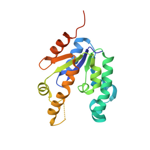
2IYR

Shikimate kinase from Mycobacterium tuberculosis in complex with shikimate


Experimental Data Snapshot

  • Method: X-RAY DIFFRACTION
  • Resolution: 1.98 Å
  • R-Value Free: 0.250 
  • R-Value Work: 0.165 
  • R-Value Observed: 0.169 

Starting Model: experimental
View more details

wwPDB Validation   3D Report Full Report


Ligand Structure Quality Assessment 


This is version 1.3 of the entry. See complete history


Literature

Mechanism of Phosphoryl Transfer Catalyzed by Shikimate Kinase from Mycobacterium Tuberculosis.

Hartmann, M.D.Bourenkov, G.P.Oberschall, A.Strizhov, N.Bartunik, H.D.

(2006) J Mol Biol 364: 411

  • DOI: https://doi.org/10.1016/j.jmb.2006.09.001
  • Primary Citation of Related Structures:  
    2IYQ, 2IYR, 2IYS, 2IYT, 2IYU, 2IYV, 2IYW, 2IYX, 2IYY, 2IYZ

  • PubMed Abstract: 

    The structural mechanism of the catalytic functioning of shikimate kinase from Mycobacterium tuberculosis was investigated on the basis of a series of high-resolution crystal structures corresponding to individual steps in the enzymatic reaction. The catalytic turnover of shikimate and ATP into the products shikimate-3-phosphate and ADP, followed by release of ADP, was studied in the crystalline environment. Based on a comparison of the structural states before initiation of the reaction and immediately after the catalytic step, we derived a structural model of the transition state that suggests that phosphoryl transfer proceeds with inversion by an in-line associative mechanism. The random sequential binding of shikimate and nucleotides is associated with domain movements. We identified a synergic mechanism by which binding of the first substrate may enhance the affinity for the second substrate.


  • Organizational Affiliation

    Max Planck Unit for Structural Molecular Biology, MPG-ASMB c/o DESY, Notkestrasse 85, 22603 Hamburg, Germany.


Macromolecules
Find similar proteins by:  (by identity cutoff)  |  3D Structure
Entity ID: 1
MoleculeChains Sequence LengthOrganismDetailsImage
SHIKIMATE KINASE
A, B
184Mycobacterium tuberculosis H37RvMutation(s): 0 
EC: 2.7.1.71
UniProt
Find proteins for P9WPY3 (Mycobacterium tuberculosis (strain ATCC 25618 / H37Rv))
Explore P9WPY3 
Go to UniProtKB:  P9WPY3
Entity Groups  
Sequence Clusters30% Identity50% Identity70% Identity90% Identity95% Identity100% Identity
UniProt GroupP9WPY3
Sequence Annotations
Expand
  • Reference Sequence
Small Molecules
Experimental Data & Validation

Experimental Data

  • Method: X-RAY DIFFRACTION
  • Resolution: 1.98 Å
  • R-Value Free: 0.250 
  • R-Value Work: 0.165 
  • R-Value Observed: 0.169 
  • Space Group: P 1
Unit Cell:
Length ( Å )Angle ( ˚ )
a = 44.444α = 90
b = 41.836β = 113.99
c = 44.408γ = 90
Software Package:
Software NamePurpose
REFMACrefinement
DENZOdata reduction
SCALEPACKdata scaling
MOLREPphasing

Structure Validation

View Full Validation Report


Ligand Structure Quality Assessment 


Entry History 

Deposition Data

Revision History  (Full details and data files)

  • Version 1.0: 2006-10-11
    Type: Initial release
  • Version 1.1: 2011-05-08
    Changes: Version format compliance
  • Version 1.2: 2011-07-13
    Changes: Version format compliance
  • Version 1.3: 2023-12-13
    Changes: Data collection, Database references, Derived calculations, Other, Refinement description