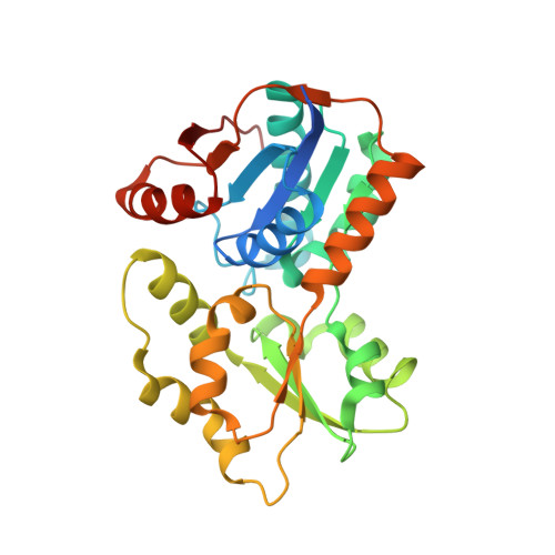
2JFN

Crystal structure of Escherichia coli glutamate racemase in complex with L- Glutamate and activator UDP-MurNAc-ala


Experimental Data Snapshot

  • Method: X-RAY DIFFRACTION
  • Resolution: 1.90 Å
  • R-Value Free: 0.244 
  • R-Value Work: 0.218 

wwPDB Validation   3D Report Full Report


Ligand Structure Quality Assessment 


This is version 1.3 of the entry. See complete history


Literature

Exploitation of Structural and Regulatory Diversity in Glutamate Racemases

Lundqvist, T.Fisher, S.L.Kern, G.Folmer, R.H.A.Xue, Y.Newton, D.T.Keating, T.A.Alm, R.A.De Jonge, B.L.M.

(2007) Nature 447: 817

  • DOI: https://doi.org/10.1038/nature05689

  • PubMed Abstract: 

    Glutamate racemase is an enzyme essential to the bacterial cell wall biosynthesis pathway, and has therefore been considered as a target for antibacterial drug discovery. We characterized the glutamate racemases of several pathogenic bacteria using structural and biochemical approaches. Here we describe three distinct mechanisms of regulation for the family of glutamate racemases: allosteric activation by metabolic precursors, kinetic regulation through substrate inhibition, and D-glutamate recycling using a d-amino acid transaminase. In a search for selective inhibitors, we identified a series of uncompetitive inhibitors specifically targeting Helicobacter pylori glutamate racemase that bind to a cryptic allosteric site, and used these inhibitors to probe the mechanistic and dynamic features of the enzyme. These structural, kinetic and mutational studies provide insight into the physiological regulation of these essential enzymes and provide a basis for designing narrow-spectrum antimicrobial agents.


  • Organizational Affiliation

    AstraZeneca Global Structural Chemistry, AstraZeneca R&D Mölndal, SE-431 83, Mölndal, Sweden.


Macromolecules
Find similar proteins by:  (by identity cutoff)  |  3D Structure
Entity ID: 1
MoleculeChains Sequence LengthOrganismDetailsImage
GLUTAMATE RACEMASE285Escherichia coliMutation(s): 0 
EC: 5.1.1.3
UniProt
Find proteins for P22634 (Escherichia coli (strain K12))
Explore P22634 
Go to UniProtKB:  P22634
Entity Groups  
Sequence Clusters30% Identity50% Identity70% Identity90% Identity95% Identity100% Identity
UniProt GroupP22634
Sequence Annotations
Expand
  • Reference Sequence
Small Molecules
Ligands 2 Unique
IDChains Name / Formula / InChI Key2D Diagram3D Interactions
UMA
Query on UMA

Download Ideal Coordinates CCD File 
C [auth A]URIDINE-5'-DIPHOSPHATE-N-ACETYLMURAMOYL-L-ALANINE
C23 H36 N4 O20 P2
NTMMCWJNQNKACG-KBKUWGQMSA-N
GLU
Query on GLU

Download Ideal Coordinates CCD File 
B [auth A]GLUTAMIC ACID
C5 H9 N O4
WHUUTDBJXJRKMK-VKHMYHEASA-N
Experimental Data & Validation

Experimental Data

  • Method: X-RAY DIFFRACTION
  • Resolution: 1.90 Å
  • R-Value Free: 0.244 
  • R-Value Work: 0.218 
  • Space Group: C 2 2 21
Unit Cell:
Length ( Å )Angle ( ˚ )
a = 83.05α = 90
b = 112.82β = 90
c = 74.12γ = 90
Software Package:
Software NamePurpose
CNXrefinement
MOSFLMdata reduction
SCALAdata scaling
SOLVEphasing

Structure Validation

View Full Validation Report


Ligand Structure Quality Assessment 


Entry History 

Deposition Data

Revision History  (Full details and data files)

  • Version 1.0: 2007-07-03
    Type: Initial release
  • Version 1.1: 2011-05-08
    Changes: Version format compliance
  • Version 1.2: 2011-07-13
    Changes: Version format compliance
  • Version 1.3: 2024-05-08
    Changes: Data collection, Database references, Other