2JI6

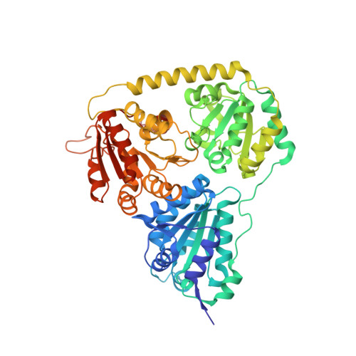
X-ray structure of Oxalyl-CoA decarboxylase in complex with 3-deaza- ThDP and oxalyl-CoA


Experimental Data Snapshot

  • Method: X-RAY DIFFRACTION
  • Resolution: 2.06 Å
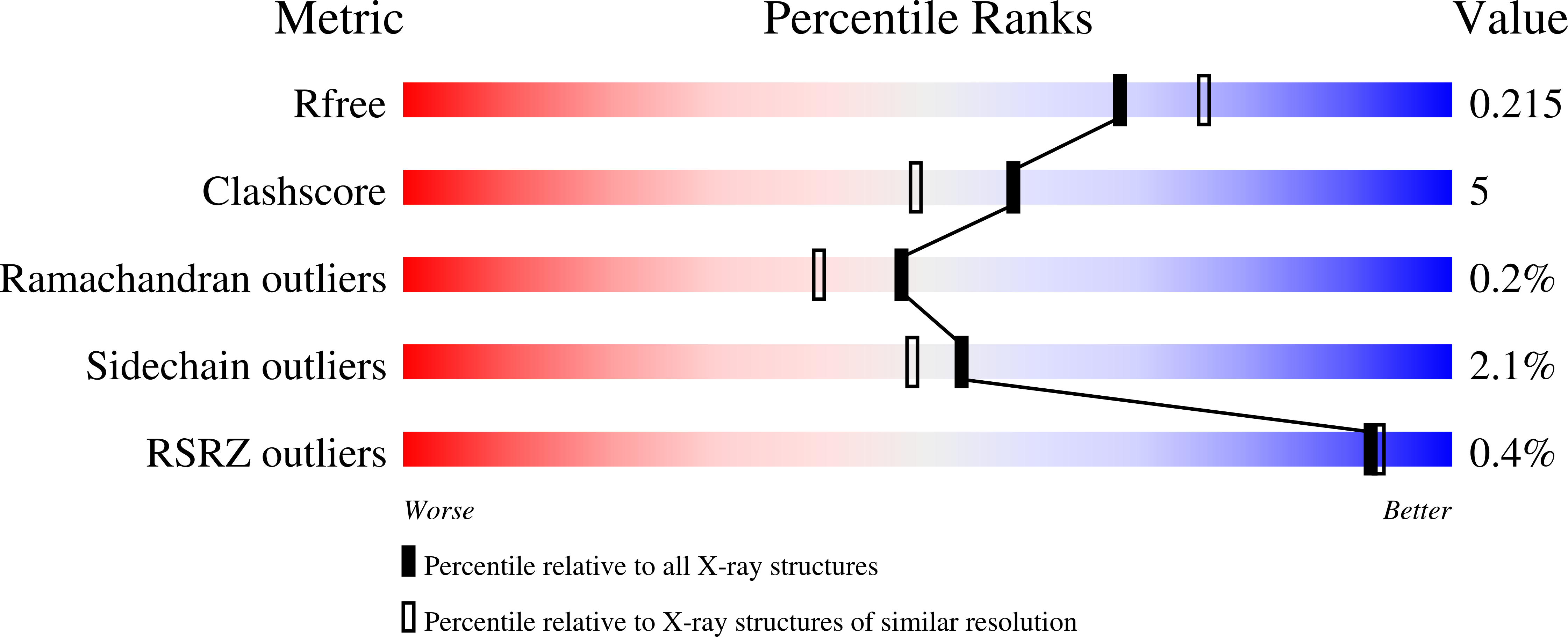
  • R-Value Free: 0.212 
  • R-Value Work: 0.174 
  • R-Value Observed: 0.176 

Starting Model: experimental
View more details

wwPDB Validation   3D Report Full Report


Ligand Structure Quality Assessment 


This is version 1.3 of the entry. See complete history


Literature

Crystallographic Snapshots of Oxalyl-Coa Decarboxylase Give Insights Into Catalysis by Nonoxidative Thdp-Dependent Decarboxylases

Berthold, C.L.Toyota, C.G.Moussatche, P.Wood, M.D.Leeper, F.Richards, N.G.J.Lindqvist, Y.

(2007) Structure 15: 853

  • DOI: https://doi.org/10.1016/j.str.2007.06.001
  • Primary Citation of Related Structures:  
    2JI6, 2JI7, 2JI8, 2JI9, 2JIB

  • PubMed Abstract: 

    Despite more than five decades of extensive studies of thiamin diphosphate (ThDP) enzymes, there remain many uncertainties as to how these enzymes achieve their rate enhancements. Here, we present a clear picture of catalysis for the simple nonoxidative decarboxylase, oxalyl-coenzyme A (CoA) decarboxylase, based on crystallographic snapshots along the catalytic cycle and kinetic data on active site mutants. First, we provide crystallographic evidence that, upon binding of oxalyl-CoA, the C-terminal 13 residues fold over the substrate, aligning the substrate alpha-carbon for attack by the ThDP-C2 atom. The second structure presented shows a covalent reaction intermediate after decarboxylation, interpreted as being nonplanar. Finally, the structure of a product complex is presented. In accordance with mutagenesis data, no side chains of the enzyme are implied to directly participate in proton transfer except the glutamic acid (Glu-56), which promotes formation of the 1',4'-iminopyrimidine tautomer of ThDP needed for activation.


  • Organizational Affiliation

    Department of Medical Biochemistry and Biophysics, Molecular Structural Biology, Karolinska Institutet, S-17177 Stockholm, Sweden.


Macromolecules
Find similar proteins by:  (by identity cutoff)  |  3D Structure
Entity ID: 1
MoleculeChains Sequence LengthOrganismDetailsImage
OXALYL-COA DECARBOXYLASE
A, B
568Oxalobacter formigenesMutation(s): 0 
EC: 4.1.1.8
UniProt
Find proteins for P40149 (Oxalobacter formigenes)
Explore P40149 
Go to UniProtKB:  P40149
Entity Groups  
Sequence Clusters30% Identity50% Identity70% Identity90% Identity95% Identity100% Identity
UniProt GroupP40149
Sequence Annotations
Expand
  • Reference Sequence
Small Molecules
Ligands 6 Unique
IDChains Name / Formula / InChI Key2D Diagram3D Interactions
OXK
Query on OXK

Download Ideal Coordinates CCD File 
F [auth A],
L [auth B]
OXALYL-COENZYME A
C23 H36 N7 O19 P3 S
QVXMZFTWJVBUHP-IBOSZNHHSA-N
ADP
Query on ADP

Download Ideal Coordinates CCD File 
E [auth A],
K [auth B]
ADENOSINE-5'-DIPHOSPHATE
C10 H15 N5 O10 P2
XTWYTFMLZFPYCI-KQYNXXCUSA-N
TPW
Query on TPW

Download Ideal Coordinates CCD File 
C [auth A],
I [auth B]
2-{4-[(4-AMINO-2-METHYLPYRIMIDIN-5-YL)METHYL]-3-METHYLTHIOPHEN-2-YL}ETHYL TRIHYDROGEN DIPHOSPHATE
C13 H19 N3 O7 P2 S
IOGGWTLVIZLGGZ-UHFFFAOYSA-N
B3P
Query on B3P

Download Ideal Coordinates CCD File 
H [auth A],
N [auth B]
2-[3-(2-HYDROXY-1,1-DIHYDROXYMETHYL-ETHYLAMINO)-PROPYLAMINO]-2-HYDROXYMETHYL-PROPANE-1,3-DIOL
C11 H26 N2 O6
HHKZCCWKTZRCCL-UHFFFAOYSA-N
PGE
Query on PGE

Download Ideal Coordinates CCD File 
G [auth A],
M [auth B]
TRIETHYLENE GLYCOL
C6 H14 O4
ZIBGPFATKBEMQZ-UHFFFAOYSA-N
MG
Query on MG

Download Ideal Coordinates CCD File 
D [auth A],
J [auth B]
MAGNESIUM ION
Mg
JLVVSXFLKOJNIY-UHFFFAOYSA-N
Experimental Data & Validation

Experimental Data

  • Method: X-RAY DIFFRACTION
  • Resolution: 2.06 Å
  • R-Value Free: 0.212 
  • R-Value Work: 0.174 
  • R-Value Observed: 0.176 
  • Space Group: P 31 2 1
Unit Cell:
Length ( Å )Angle ( ˚ )
a = 127.002α = 90
b = 127.002β = 90
c = 151.813γ = 120
Software Package:
Software NamePurpose
REFMACrefinement
MOSFLMdata reduction
SCALAdata scaling
MOLREPphasing

Structure Validation

View Full Validation Report


Ligand Structure Quality Assessment 


Entry History 

Deposition Data

Revision History  (Full details and data files)

  • Version 1.0: 2007-07-17
    Type: Initial release
  • Version 1.1: 2011-07-13
    Changes: Advisory, Version format compliance
  • Version 1.2: 2018-01-17
    Changes: Data collection
  • Version 1.3: 2023-12-13
    Changes: Data collection, Database references, Derived calculations, Other, Refinement description