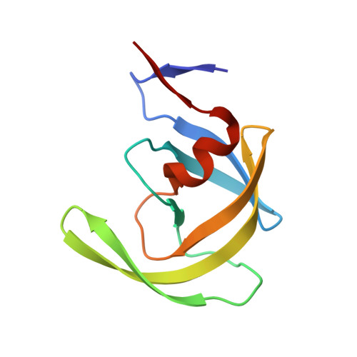
2QHC

The Influence of I47A Mutation on Reduced Susceptibility to the Protease Inhibitor Lopinavir


Experimental Data Snapshot

  • Method: X-RAY DIFFRACTION
  • Resolution: 2.80 Å
  • R-Value Free: 0.228 
  • R-Value Work: 0.191 
  • R-Value Observed: 0.193 

Starting Model: experimental
View more details

wwPDB Validation   3D Report Full Report


Ligand Structure Quality Assessment 


This is version 1.3 of the entry. See complete history


Literature

Enzymatic and structural analysis of the I47A mutation contributing to the reduced susceptibility to HIV protease inhibitor lopinavir.

Saskova, K.G.Kozisek, M.Lepsik, M.Brynda, J.Rezacova, P.Vaclavikova, J.Kagan, R.M.Machala, L.Konvalinka, J.

(2008) Protein Sci 17: 1555-1564

  • DOI: https://doi.org/10.1110/ps.036079.108
  • Primary Citation of Related Structures:  
    2QHC, 2Z54

  • PubMed Abstract: 

    Lopinavir (LPV) is a second-generation HIV protease inhibitor (PI) designed to overcome resistance development in patients undergoing long-term antiviral therapy. The mutation of isoleucine at position 47 of the HIV protease (PR) to alanine is associated with a high level of resistance to LPV. In this study, we show that recombinant PR containing a single I47A substitution has the inhibition constant (K(i) ) value for lopinavir by two orders of magnitude higher than for the wild-type PR. The addition of the I47A substitution to the background of a multiply mutated PR species from an AIDS patient showed a three-order-of-magnitude increase in K(i) in vitro relative to the patient PR without the I47A mutation. The crystal structure of I47A PR in complex with LPV showed the loss of van der Waals interactions in the S2/S2' subsites. This is caused by the loss of three side-chain methyl groups due to the I47A substitution and by structural changes in the A47 main chain that lead to structural changes in the flap antiparallel beta-strand. Furthermore, we analyzed possible interaction of the I47A mutation with secondary mutations V32I and I54V. We show that both mutations in combination with I47A synergistically increase the relative resistance to LPV in vitro. The crystal structure of the I47A/I54V PR double mutant in complex with LPV shows that the I54V mutation leads to a compaction of the flap, and molecular modeling suggests that the introduction of the I54V mutation indirectly affects the strain of the bound inhibitor in the PR binding cleft.


  • Organizational Affiliation

    Gilead Sciences and IOCB Research Center, Institute of Organic Chemistry and Biochemistry, Academy of Sciences of the Czech Republic, Prague 6, Czech Republic.


Macromolecules
Find similar proteins by:  (by identity cutoff)  |  3D Structure
Entity ID: 1
MoleculeChains Sequence LengthOrganismDetailsImage
PROTEASE RETROPEPSIN
A, B
99Human immunodeficiency virus 1Mutation(s): 1 
Gene Names: gag-pol
EC: 3.4.23.16
UniProt
Find proteins for Q9J2P7 (Human immunodeficiency virus 1)
Explore Q9J2P7 
Go to UniProtKB:  Q9J2P7
Entity Groups  
Sequence Clusters30% Identity50% Identity70% Identity90% Identity95% Identity100% Identity
UniProt GroupQ9J2P7
Sequence Annotations
Expand
  • Reference Sequence
Small Molecules
Ligands 2 Unique
IDChains Name / Formula / InChI Key2D Diagram3D Interactions
AB1
Query on AB1

Download Ideal Coordinates CCD File 
E [auth B]N-{1-BENZYL-4-[2-(2,6-DIMETHYL-PHENOXY)-ACETYLAMINO]-3-HYDROXY-5-PHENYL-PENTYL}-3-METHYL-2-(2-OXO-TETRAHYDRO-PYRIMIDIN-1-YL)-BUTYRAMIDE
C37 H48 N4 O5
KJHKTHWMRKYKJE-SUGCFTRWSA-N
BME
Query on BME

Download Ideal Coordinates CCD File 
C [auth A],
D [auth B]
BETA-MERCAPTOETHANOL
C2 H6 O S
DGVVWUTYPXICAM-UHFFFAOYSA-N
Binding Affinity Annotations 
IDSourceBinding Affinity
AB1 BindingDB:  2QHC Ki: min: 5.00e-3, max: 2.4 (nM) from 6 assay(s)
IC50: 25 (nM) from 1 assay(s)
PDBBind:  2QHC Ki: 1.1 (nM) from 1 assay(s)
Experimental Data & Validation

Experimental Data

  • Method: X-RAY DIFFRACTION
  • Resolution: 2.80 Å
  • R-Value Free: 0.228 
  • R-Value Work: 0.191 
  • R-Value Observed: 0.193 
  • Space Group: P 61
Unit Cell:
Length ( Å )Angle ( ˚ )
a = 62.576α = 90
b = 62.576β = 90
c = 81.934γ = 120
Software Package:
Software NamePurpose
MAR345dtbdata collection
MOLREPphasing
REFMACrefinement
MOSFLMdata reduction
SCALAdata scaling

Structure Validation

View Full Validation Report


Ligand Structure Quality Assessment 


Entry History 

Deposition Data

Revision History  (Full details and data files)

  • Version 1.0: 2008-07-22
    Type: Initial release
  • Version 1.1: 2011-07-13
    Changes: Version format compliance
  • Version 1.2: 2021-10-20
    Changes: Database references, Derived calculations, Structure summary
  • Version 1.3: 2023-08-30
    Changes: Data collection, Refinement description